The orbital floor is designed to break. It gives way so that the pressure of a blow is dissipated downward instead of rupturing the globe.
An orbital blowout fracture is that protective mechanism doing its job – and then causing its own problems.
Orbital fat and muscle can herniate into the sinus below, the eye can sink backward, and eye movements can become restricted.
Most of these fractures do not need surgery, and many that look dramatic settle with time.
But one paediatric variant, in which a green-stick fracture traps the inferior rectus, is a surgical emergency measured in hours because the muscle is being strangled.
Knowing which fracture can wait, which needs planned repair, and which cannot wait at all is the core of managing orbital trauma.
What Is an Orbital Blowout Fracture?
An orbital blowout fracture is a fracture of the orbital walls – most often the floor or medial wall – caused by blunt trauma, with the orbital rim left intact.
The patterns that matter are:
- Orbital floor fracture – the commonest, with herniation of orbital contents into the maxillary sinus
- Medial wall fracture – through the thin lamina papyracea into the ethmoid sinus, often with orbital emphysema
- Combined floor and medial wall fracture – larger defects with a higher risk of significant enophthalmos
- Trapdoor (white-eyed) fracture – a paediatric green-stick fracture that springs shut and traps tissue, with minimal external signs
- Orbital roof fracture – less common, more frequent in children, and associated with intracranial injury
The distinction between an adult comminuted floor fracture and a paediatric trapdoor fracture is the single most consequential one in this whole topic.
Epidemiology
Orbital fractures follow the epidemiology of facial trauma generally.
- Assault, sports injury, falls and road traffic collisions account for the majority
- Young adult men are disproportionately represented
- The orbital floor and medial wall are the commonest sites because they are the thinnest orbital walls
- Trapdoor fractures occur predominantly in children and adolescents, whose bone is more elastic and tends to hinge rather than shatter
- Associated ocular injury is common enough that a full eye examination is mandatory in every case
A significant proportion of patients with an orbital fracture have a concurrent ocular injury, so the fracture must never be treated as the whole diagnosis.
All-fit Slit-Lamp Adapter
Record and share exactly what you see at the slit lamp. One adapter fits any slit lamp or surgical microscope — and any smartphone.
From Choroida — the team behind this sitePathophysiology
Two mechanisms are described, and they are not mutually exclusive.
- The hydraulic theory – a blow to the globe raises intraorbital pressure, which is transmitted to the walls and fractures the weakest point
- The buckling theory – force applied to the orbital rim transmits a buckling wave through the floor, fracturing it without direct globe compression
- Orbital fat, and sometimes the inferior rectus muscle, prolapses through the defect into the sinus below
- Loss of orbital volume support and post-traumatic fat atrophy cause the globe to sink backward and downward – enophthalmos and hypoglobus
- Entrapment or tethering of tissue restricts eye movement and produces diplopia
- In trapdoor fractures the bone recoils and clamps the entrapped muscle, compromising its blood supply and causing ischaemia
Ischaemia is precisely why the trapdoor variant is urgent: this is a strangulated muscle, and the clock runs in hours rather than weeks.
Clinical Presentation
Symptoms
- Periorbital swelling, bruising and pain following blunt trauma
- Double vision, characteristically worse on upgaze with a floor fracture
- Numbness of the cheek, upper lip, upper teeth and gum from infraorbital nerve injury
- Orbital emphysema with crepitus, classically worse after nose-blowing in medial wall fractures
- A sunken appearance of the eye, which often becomes apparent only as swelling settles
- Nausea, vomiting and bradycardia – the oculocardiac reflex, a red flag for muscle entrapment
Examination Findings
- Restricted eye movement, particularly limitation of upgaze, with diplopia in that direction
- Enophthalmos measured by exophthalmometry, often masked initially by orbital swelling
- Infraorbital hypoaesthesia mapped over the cheek and upper lip
- Subcutaneous emphysema and crepitus on gentle palpation
- A forced duction test to distinguish true mechanical restriction from a neurogenic palsy or pain-limited movement
- Full ophthalmic assessment – visual acuity, pupils, intraocular pressure, and dilated fundus examination for hyphema, retinal dialysis, commotio retinae or globe rupture
In children, the absence of bruising is misleading rather than reassuring – the “white-eyed blowout fracture” describes a quiet-looking eye with a trapped muscle underneath.
Diagnostic Evaluation
Imaging
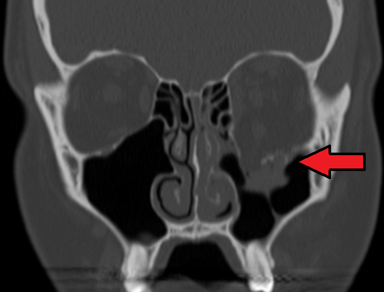
- CT of the orbits with fine cuts in axial and coronal planes is the investigation of choice
- Coronal images best demonstrate floor defects, herniated contents and the position of the inferior rectus
- A rounded inferior rectus on coronal imaging suggests entrapment rather than simple prolapse
- CT also identifies associated facial fractures and intraorbital foreign bodies
Ophthalmic Assessment
- Complete examination to exclude concurrent globe injury, which changes management priorities entirely
- Orthoptic assessment with formal measurement of the deviation and field of binocular single vision
- Serial assessment as swelling resolves, since both diplopia and enophthalmos evolve over the first two weeks
Identifying the Emergency
- A child or young adult with marked motility restriction, little swelling, pain on eye movement, and nausea or bradycardia
- This combination should prompt immediate imaging and same-day surgical referral
The oculocardiac reflex in a young patient after orbital trauma is one of the few findings in ophthalmology that should move a patient straight to theatre.
Differential Diagnosis
Causes of restricted movement and diplopia after orbital trauma include:
- Mechanical entrapment of the inferior rectus – positive forced duction test
- Orbital haemorrhage and oedema causing restriction without entrapment – improves as swelling settles
- Cranial nerve palsy from head injury – negative forced duction test
- Direct extraocular muscle contusion
- Orbital compartment syndrome with retrobulbar haemorrhage – a separate sight-threatening emergency requiring immediate canthotomy and cantholysis
- Globe rupture, which must be excluded before any forced duction testing is attempted
Forced duction testing is contraindicated until globe rupture has been excluded, since manipulating a ruptured eye can expel intraocular contents.
Management
Immediate Care
- Advise against nose-blowing, which forces air into the orbit and can cause dramatic emphysema or even orbital compartment syndrome
- Ice packs and head elevation to limit swelling
- Antibiotic cover is often given where the fracture communicates with a sinus, though practice varies
- A short course of oral corticosteroids is sometimes used to reduce oedema and clarify the true extent of restriction
Immediate Surgery
- Trapdoor fracture with muscle entrapment, particularly in children, ideally within 24 to 48 hours to prevent permanent muscle ischaemia and fibrosis
- Associated oculocardiac reflex with bradycardia, nausea and vomiting
Planned or Deferred Repair
- Repair is commonly considered within roughly two weeks for persistent symptomatic diplopia in functional gaze positions, significant enophthalmos, or a large floor defect likely to produce late enophthalmos
- Surgery involves releasing herniated tissue and reconstructing the floor with an implant
- Many small fractures with resolving diplopia and no significant enophthalmos are managed conservatively with observation
- Delayed repair is more difficult once scarring has occurred, which is why the two-week window matters
The practical rule is that most adult fractures can be observed for a fortnight while swelling settles, but a child with a trapped muscle cannot wait even a day.
Prognosis
Most patients do well, and the outlook is best when the urgent cases are identified early.
- Diplopia from swelling and haemorrhage usually resolves over days to weeks without surgery
- Trapdoor fractures released promptly generally recover full motility; delayed release risks permanent restriction from muscle fibrosis
- Infraorbital numbness improves in many patients over months, but can be permanent
- Enophthalmos may become apparent only after swelling resolves, and is harder to correct once late
- Persistent diplopia in primary or reading position after adequate repair may require prisms or strabismus surgery
Because both diplopia and enophthalmos declare themselves as swelling settles, review at one to two weeks is what turns an initial assessment into a reliable one.


Document what you see
Two smartphone imaging tools built for everyday clinic use — one for the slit lamp, one for the fundus.
From Choroida — the team behind this siteReferences
- Burnstine MA. Clinical recommendations for repair of isolated orbital floor fractures: an evidence-based analysis. Ophthalmology. 2002.
- Jordan DR, Allen LH, White J, et al. Intervention within days for some orbital floor fractures: the white-eyed blowout. Ophthalmic Plastic and Reconstructive Surgery. 1998.
- Boyette JR, Pemberton JD, Bonilla-Velez J. Management of orbital fractures: challenges and solutions. Clinical Ophthalmology. 2015.
- Orbital Floor Fracture. StatPearls, NCBI Bookshelf.
- Orbital Fractures. EyeWiki, American Academy of Ophthalmology.
Test yourself
A few questions straight from this article.
-
What defines an orbital blowout fracture?
A blowout fracture involves the orbital walls, usually the floor or medial wall, from blunt trauma, while the orbital rim itself remains intact. -
What does the buckling theory propose as the mechanism of an orbital floor fracture?
The buckling theory holds that force applied to the rim transmits a buckling wave through the floor without direct globe compression, complementing the hydraulic theory. -
Why is a paediatric trapdoor orbital fracture a surgical emergency?
In a trapdoor green-stick fracture the bone springs shut on the entrapped muscle and compromises its blood supply, so the muscle is effectively strangled and the clock runs in hours. -
Which combination after orbital trauma should move a young patient straight to theatre?
That combination is the oculocardiac reflex of a white-eyed trapdoor fracture with muscle entrapment, and it warrants immediate imaging and same-day surgery. -
Which CT appearance of the inferior rectus suggests entrapment rather than simple prolapse?
Fine-cut coronal CT is the investigation of choice, and a rounded inferior rectus on those images suggests entrapment rather than simple prolapse of orbital contents. -
Why must globe rupture be excluded before a forced duction test is performed?
Forced duction testing distinguishes mechanical restriction from a neurogenic palsy, but it is contraindicated until rupture is excluded because manipulation can expel intraocular contents. -
Injury to which nerve explains numbness of the cheek, upper lip and upper teeth after a floor fracture?
Infraorbital nerve injury within the floor produces hypoaesthesia mapped over the cheek, upper lip, upper teeth and gum, and it may improve over months or be permanent. -
Why are patients with an orbital blowout fracture told not to blow their nose?
Nose-blowing drives sinus air into the orbit, producing dramatic emphysema and crepitus and occasionally orbital compartment syndrome, so it is advised against from the outset. -
Within what time frame should a trapdoor fracture with muscle entrapment ideally be released?
Entrapped muscle should ideally be released within 24 to 48 hours to prevent permanent ischaemia and fibrosis, whereas most adult fractures can be observed for about two weeks. -
Why is review one to two weeks after an orbital blowout fracture important?
Diplopia from swelling and haemorrhage often resolves while enophthalmos only becomes apparent as oedema subsides, so reassessment at one to two weeks makes the initial assessment reliable.

Writes and reviews the clinical articles on Educate. Every post is checked against peer-reviewed sources before it goes live.