
Case Study
A 62-year-old male presented with progressive difficulty in reading and recognizing faces over the past decade.

He reported no significant medical history but noted that his father experienced similar symptoms later in life. Ophthalmic examination revealed central scotomas and subtle pigmentary changes in the macula.
Optical coherence tomography (OCT) demonstrated thinning of the retinal pigment epithelium (RPE) and loss of choroidal structure in the central macular region.
Genetic testing confirmed a mutation in the PRPH2 gene, leading to a diagnosis of Central Areolar Choroidal Dystrophy (CACD).
Disease entity
Central Areolar Choroidal Dystrophy (CACD) is a rare inherited retinal disease characterized by progressive degeneration of the macular area, resulting in central vision loss.
CACD typically presents in middle-aged to older individuals, although early signs may appear in younger patients. It is often misdiagnosed as age-related macular degeneration (AMD) due to overlapping clinical features.
However, CACD has distinct genetic and pathophysiological characteristics that warrant separate consideration.
The condition significantly impacts the quality of life, particularly due to central scotomas affecting daily activities such as reading and driving.
Advances in genetic and imaging technologies have enhanced the understanding and diagnostic accuracy of CACD, paving the way for potential therapeutic interventions.
Fundus Explorer Pro
Photograph the retinal findings described here with the phone already in your pocket — 22 D optics and built-in illumination in one handheld unit.
From Choroida — the team behind this sitePathophysiology
CACD primarily affects the retinal pigment epithelium (RPE) and choroid, leading to progressive atrophy in the macular region.
- Genetic Mutations:
- Mutations in the PRPH2 gene (formerly RDS), responsible for encoding peripherin-2, are the most common cause of CACD.
- Other implicated genes include GUCY2D and CTNNA1, highlighting genetic heterogeneity.
- Progressive Atrophy:
- Degeneration begins with subtle changes in the RPE and progresses to complete atrophy of the RPE and choroid in the macular area.
- This results in photoreceptor dysfunction, particularly affecting cone cells.
- Secondary Choroidal Changes:
- Loss of choroidal vasculature exacerbates photoreceptor damage by disrupting nutrient and oxygen supply.
Epidemiology
CACD is a rare condition with an estimated prevalence of 1 in 50,000 individuals.
- Age of Onset: Symptoms usually manifest in the fourth to sixth decades of life, although earlier onset may occur in severe cases.
- Inheritance Pattern: Autosomal dominant inheritance is most common, but sporadic cases are also reported.
- Geographic and Ethnic Variability: CACD appears to affect individuals of all ethnicities equally, although genetic mutation frequencies may vary.
Clinical Features
The clinical progression of CACD is typically divided into four stages:
- Stage 1 (Early):
- Subtle pigmentary changes in the macula.
- Mild visual symptoms, often unnoticed by the patient.
- Stage 2 (Intermediate):
- More pronounced pigment clumping and localized RPE atrophy.
- Patients may report difficulty in low-light conditions or mild central vision disturbances.
- Stage 3 (Advanced):
- Enlargement of RPE atrophic areas.
- Central scotomas become apparent, significantly impairing vision.
- Stage 4 (Late):
- Complete atrophy of the macula, involving RPE, choroid, and photoreceptors.
- Severe central vision loss, leaving peripheral vision intact.
Common symptoms across stages include:
- Central vision loss (progressive and bilateral).
- Difficulty with reading and face recognition.
- Photophobia in some patients.
Diagnosis
Diagnosis of CACD requires a combination of clinical evaluation, imaging, and genetic testing.
- Clinical Examination:
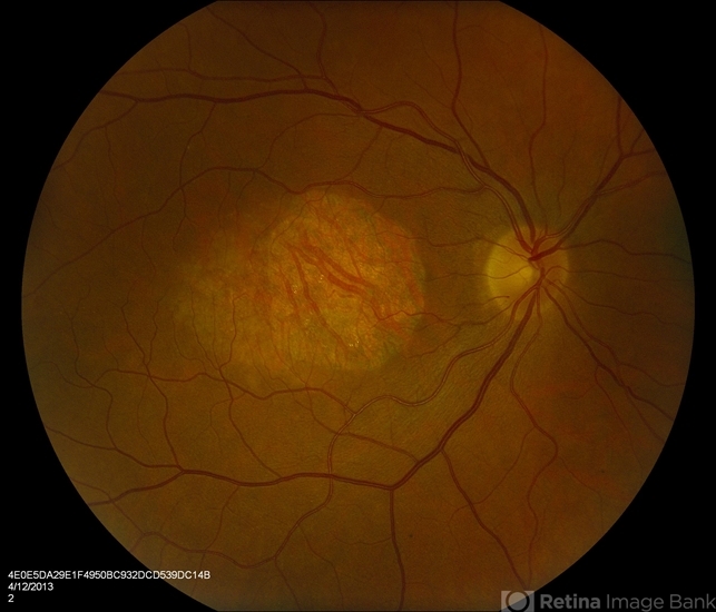
- Fundus examination reveals central macular atrophy with well-demarcated borders.
- Pigmentary changes, including clumping or mottling, may be present.
- Imaging Modalities:
- Fundus Autofluorescence (FAF): Highlights RPE loss and areas of atrophy.
- Optical Coherence Tomography (OCT): Demonstrates thinning of the RPE and loss of outer retinal layers.
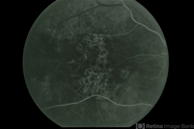
- Fluorescein Angiography (FA): Shows window defects corresponding to areas of RPE atrophy.
- Genetic Testing:
- Identifies causative mutations, particularly in the PRPH2 gene.
- Confirms diagnosis and aids in family counseling.
- Electrophysiology:
- Electroretinography (ERG) may show reduced cone function, particularly in advanced stages.
Differential Diagnosis
CACD must be differentiated from other conditions affecting the macula, including:
- Age-Related Macular Degeneration (AMD): The presence of drusen in AMD helps differentiate it from CACD.
- Stargardt Disease: Earlier onset and a flecked fundus pattern distinguish Stargardt disease.
- Pattern Dystrophies: Characterized by a “butterfly” or other unique pigment patterns.
Management
Currently, there is no cure for CACD, and management focuses on supportive care and low-vision rehabilitation.
- Low-Vision Aids:
- Magnifiers, electronic reading devices, and adaptive technologies enhance visual function.
- Photoprotection:
- Sunglasses with UV protection may help slow disease progression by reducing phototoxic damage.
- Nutritional Support:
- Antioxidant supplementation, though unproven in CACD, may offer some benefits based on evidence from AMD studies.
- Emerging Therapies:
- Gene Therapy: Experimental approaches targeting specific genetic mutations.
- Cell-Based Therapies: RPE transplantation or stem cell-derived photoreceptor implants.
- Pharmacologic Interventions: Neuroprotective agents and drugs targeting choroidal blood flow are under investigation.
- Monitoring:
Regular follow-ups to monitor disease progression and adapt management strategies.
Prognosis
The prognosis of CACD depends on the stage of disease at diagnosis and the rate of progression:
- Early Diagnosis: This may allow interventions to slow progression and preserve functional vision.
- Advanced Stages: Central vision is severely affected, leading to significant impairment in daily life activities.
While CACD does not affect peripheral vision, central vision loss remains irreversible.

Prevention
Preventive strategies for CACD are limited due to its genetic nature. However, early detection in families with a history of CACD can facilitate monitoring and timely interventions.
Genetic counseling plays a critical role in identifying at-risk individuals and guiding family planning decisions.


Document what you see
Two smartphone imaging tools built for everyday clinic use — one for the slit lamp, one for the fundus.
From Choroida — the team behind this siteReferences
- Allikmets R, Singh N, Sun H, et al. Genetic insights into central areolar choroidal dystrophy. Prog Retin Eye Res. 2020;45:1-14.
- Rosenberg T, Schwartz M. Autosomal dominant central areolar choroidal dystrophy: Clinical and genetic findings. Br J Ophthalmol. 2006;90(12):1457-1461.
- Parmeggiani F, Sodi A, Campa C, et al. CACD imaging and electrophysiology: Advances in diagnosis. Eye (Lond). 2019;33(3):381-388.
- Birtel J, Gliem M, Herrmann P, et al. Inherited macular and choroidal dystrophies: Diagnostic and therapeutic advances. Ophthalmology. 2021;128(7):997-1010.
Test yourself
A few questions straight from this article.
-
Which gene mutation most commonly causes central areolar choroidal dystrophy (CACD)?
Mutations in PRPH2, formerly called RDS and encoding peripherin-2, are the most common cause of CACD; GUCY2D and CTNNA1 are also implicated. -
What is the estimated prevalence of central areolar choroidal dystrophy?
CACD is rare, with an estimated prevalence of about 1 in 50,000 individuals, symptoms usually appearing in the fourth to sixth decades. -
Which inheritance pattern is most common in central areolar choroidal dystrophy?
Autosomal dominant inheritance is most common in CACD, although sporadic cases are also reported. -
How many clinical stages is central areolar choroidal dystrophy divided into?
The article divides CACD into four stages, from subtle macular pigmentary change through to complete macular atrophy with severe central vision loss. -
What characterises stage 4 (late) central areolar choroidal dystrophy?
Stage 4 involves complete atrophy of the macula including RPE, choroid and photoreceptors, causing severe central vision loss while peripheral vision remains intact. -
Which fundus appearance is characteristic of central areolar choroidal dystrophy?
Fundus examination shows central macular atrophy with well-demarcated borders, sometimes with pigment clumping or mottling. -
What does fluorescein angiography show in central areolar choroidal dystrophy?
Fluorescein angiography shows window defects corresponding to areas of RPE atrophy, while autofluorescence highlights RPE loss and OCT shows outer retinal thinning. -
Which feature best separates age-related macular degeneration from central areolar choroidal dystrophy?
CACD is often misdiagnosed as AMD, but the presence of drusen in AMD is what helps differentiate the two conditions. -
Which electrophysiology finding is expected in advanced central areolar choroidal dystrophy?
Electroretinography may show reduced cone function, particularly in advanced stages, reflecting the cone-predominant photoreceptor dysfunction of CACD. -
What does current management of central areolar choroidal dystrophy consist of?
There is no cure; care centres on low-vision aids, UV-protective sunglasses, nutritional support and regular follow-up, with gene and cell-based therapies still experimental.
