
For years, a diagnosis of geographic atrophy came with a specific, unwelcome kind of honesty: there was nothing to offer.
Anti-VEGF drugs treated the wet form of macular degeneration. The dry, atrophic form simply progressed.
Geographic atrophy is the advanced form of dry age-related macular degeneration, and it is now, for the first time, a treatable disease.
Two complement inhibitors have reached approval on the strength of trials that slowed the rate of atrophy expansion.
What they have not done – and this distinction matters enormously for how the treatment is discussed with patients – is restore vision already lost.
What Is Geographic Atrophy?
Geographic atrophy (GA) is a sharply demarcated area of loss of the retinal pigment epithelium, photoreceptors and choriocapillaris, representing the end-stage of dry age-related macular degeneration.
Two treatment-relevant features define the modern approach:
- Lesions grow over time, with growth rate influenced by lesion size, location, multifocality and genetic factors
- Once retinal tissue within a lesion is atrophic, that vision loss is permanent – treatment targets future growth, not existing damage
This second point is the single most important thing to communicate to a newly diagnosed patient, because it reframes what “treatment” can realistically deliver.
Epidemiology
Geographic atrophy is a major and growing cause of irreversible vision loss.
- It affects a substantial proportion of patients with advanced age-related macular degeneration, and prevalence rises steeply with age
- Bilateral involvement is common, though the two eyes often progress at different rates
- Risk factors mirror those for AMD generally – age, smoking, family history and specific genetic variants including complement factor H and ARMS2
- As populations age, geographic atrophy prevalence is projected to rise substantially over the coming decades
The scale of the unmet need is exactly why complement inhibition attracted such sustained investment despite a difficult trial history.
Fundus Explorer Pro
Photograph the retinal findings described here with the phone already in your pocket — 22 D optics and built-in illumination in one handheld unit.
From Choroida — the team behind this sitePathophysiology
The complement system is now understood to be central to how atrophy develops and spreads.
- Chronic low-grade inflammation and complement activation within the retinal pigment epithelium and choriocapillaris drive progressive cell loss
- Drusen and complement deposits accumulate at the level of the RPE and Bruch membrane over years before atrophy becomes clinically apparent
- Loss of RPE support leads to photoreceptor death, and loss of the choriocapillaris compounds the ischaemic insult
- The alternative complement pathway, and specifically complement component 3 and complement factor C5, have emerged as the principal therapeutic targets
Targeting complement rather than VEGF reflects a fundamentally different disease mechanism – this is a chronic inflammatory and degenerative process, not a vascular leak.
Clinical Presentation
Symptoms
- Gradual, often asymmetric central vision loss, frequently first noticed as difficulty reading in dim light
- A scotoma that patients may describe as a blurred or missing patch rather than a sharp black spot
- Preserved peripheral vision even in advanced disease, since GA is a macular process
- Slow progression over years rather than the days-to-weeks course of wet AMD
Examination and Imaging Findings
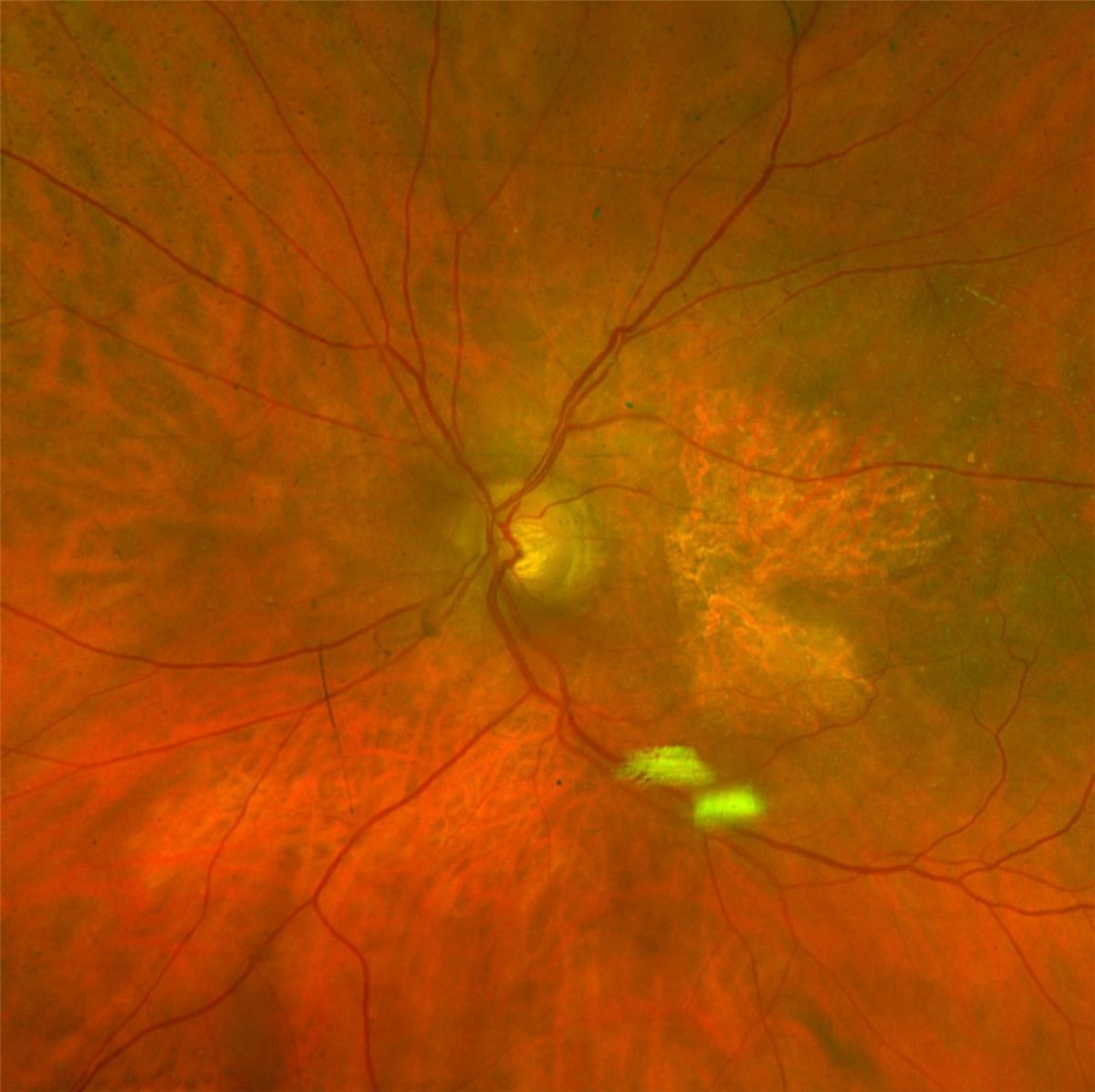
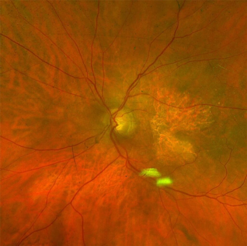
- A well-demarcated area of RPE loss on fundus examination, with underlying choroidal vessels visible through the atrophic patch
- Fundus autofluorescence showing a hypoautofluorescent lesion with a variable hyperautofluorescent junctional zone, the region felt to reflect stressed RPE at highest risk of future involvement
- OCT showing loss of the RPE band, outer retinal atrophy and increased choroidal signal transmission beneath the lesion
- Drusen and pigmentary change typically present in the surrounding retina, reflecting the underlying AMD
Multimodal imaging – autofluorescence combined with OCT – is now standard for both diagnosis and for monitoring the lesion growth that guides treatment decisions.
Diagnostic Evaluation
Establishing the Diagnosis
- Dilated fundus examination and colour fundus photography
- Fundus autofluorescence to delineate lesion borders precisely
- OCT to confirm outer retinal and RPE loss and exclude coexistent neovascular disease
Assessing Risk of Progression
- Baseline lesion size and location relative to the fovea
- Lesion configuration – multifocal lesions tend to progress faster than a single confluent lesion
- Serial imaging over time to establish an individual growth rate before considering treatment
Excluding Neovascular Conversion
- OCT and, where needed, OCT angiography to detect subclinical choroidal neovascularisation, which can develop within or adjacent to atrophic lesions and changes management substantially
Distinguishing GA from occult or non-exudative neovascular membranes matters practically, since the two are managed with entirely different drug classes.
Management
Complement Inhibitor Therapy
- Pegcetacoplan, a C3 inhibitor, and avacincaptad pegol, a C5 inhibitor, are given as monthly or bimonthly intravitreal injections
- Pivotal trials demonstrated a statistically significant reduction in the rate of lesion growth compared with sham injection, in the range of roughly 15 to 20 percent depending on the agent, regimen and follow-up duration
- Neither agent has been shown to improve visual acuity, and both carry a small but real risk of progression to neovascular AMD during treatment, requiring monitoring for conversion
- Patient selection favours lesions positioned where slowing growth is most likely to preserve functionally important vision – extrafoveal lesions with intact foveal sparing are a common target
Supportive Management
- AREDS2 formulation supplementation, which reduces progression from intermediate to advanced AMD but has not been shown to slow established GA growth
- Smoking cessation, which remains the single most impactful modifiable risk factor across the AMD spectrum
- Low vision rehabilitation and referral to visual aids services, which remain central to functional outcome regardless of drug therapy
- Monitoring of the fellow eye, since progression is frequently asymmetric
The treatment decision here is unusually dependent on shared decision-making – a monthly injection burden for a modest reduction in growth rate, with no promise of visual improvement, is a trade-off patients must weigh for themselves.
Differential Diagnosis
Conditions that can mimic geographic atrophy include:
- Pattern dystrophies, including adult-onset vitelliform lesions – typically earlier onset and different autofluorescence patterns
- Stargardt disease – earlier onset with a characteristic flecked appearance and dark choroid on fluorescein angiography
- Toxic maculopathy from hydroxychloroquine – bull’s-eye pattern with a relevant drug history
- Chloroquine or other drug-induced retinopathy
- Choroideremia and other inherited chorioretinal atrophies, distinguished by earlier onset and characteristic peripheral involvement
Age at onset and the pattern of autofluorescence are usually enough to separate age-related GA from its inherited mimics.
Prognosis
Geographic atrophy remains a progressive disease even with treatment, and expectations need to be set accordingly.
- Lesions continue to grow on complement inhibitor therapy, just more slowly than on sham treatment in trial populations
- Central vision loss eventually occurs when growth reaches the fovea, regardless of treatment, though slower growth may delay that point
- Peripheral vision is preserved throughout, and complete blindness from GA alone is rare
- Long-term real-world data on functional benefit, and on the durability of the growth-rate reduction, are still accumulating
Geographic atrophy treatment is best understood as buying time rather than reversing disease – a meaningful first step, but one that leaves plenty of room for the next generation of therapy.


Document what you see
Two smartphone imaging tools built for everyday clinic use — one for the slit lamp, one for the fundus.
From Choroida — the team behind this siteReferences
- Heier JS, Lad EM, Holz FG, et al. Pegcetacoplan for the treatment of geographic atrophy (OAKS and DERBY). The Lancet. 2023.
- Khanani AM, Patel SS, Staurenghi G, et al. Efficacy and safety of avacincaptad pegol (GATHER1, GATHER2). The Lancet. 2023.
- Age-Related Eye Disease Study 2 Research Group. Lutein, zeaxanthin, and omega-3 fatty acids for AMD (AREDS2). JAMA. 2013.
- Geographic Atrophy. EyeWiki, American Academy of Ophthalmology.
- Age-Related Macular Degeneration, Geographic Atrophy. StatPearls, NCBI Bookshelf.
Test yourself
A few questions straight from this article.
-
Geographic atrophy represents the end stage of which form of age-related macular degeneration?
Geographic atrophy is the advanced form of dry AMD, a sharply demarcated loss of retinal pigment epithelium, photoreceptors and choriocapillaris. -
Which pathway has emerged as the principal therapeutic target in geographic atrophy?
Chronic complement activation drives RPE and choriocapillaris loss, and the alternative pathway, specifically C3 and C5, provides the therapeutic targets. -
What should a patient newly diagnosed with geographic atrophy be told that treatment can achieve?
Vision lost within an atrophic lesion is permanent; complement inhibitors target future growth rather than existing damage, and neither agent improves acuity. -
By roughly how much did pivotal trials show complement inhibitors reduce geographic atrophy lesion growth versus sham?
Pivotal trials demonstrated a statistically significant reduction in lesion growth rate of roughly 15 to 20 percent, varying with agent, regimen and follow-up duration. -
On fundus autofluorescence in geographic atrophy, what does the junctional zone around the lesion show?
The lesion itself is hypoautofluorescent, while a variable hyperautofluorescent junctional zone marks stressed RPE thought to be at highest risk of future involvement. -
Which lesion configuration in geographic atrophy is associated with faster progression?
When assessing progression risk, multifocal lesions tend to progress faster than a single confluent lesion, alongside baseline size and location relative to the fovea. -
Which feature best separates Stargardt disease from age-related geographic atrophy?
Stargardt disease presents earlier with a characteristic flecked appearance and a dark choroid on fluorescein angiography, unlike age-related geographic atrophy. -
How are pegcetacoplan and avacincaptad pegol administered in geographic atrophy?
Pegcetacoplan, a C3 inhibitor, and avacincaptad pegol, a C5 inhibitor, are both given as monthly or bimonthly intravitreal injections. -
Which risk during complement inhibitor therapy for geographic atrophy requires ongoing monitoring?
Both approved agents carry a small but real risk of conversion to neovascular AMD during treatment, so patients must be monitored for that conversion. -
What happens to peripheral vision over the course of geographic atrophy?
Geographic atrophy is a macular process, so peripheral vision is preserved even in advanced disease and complete blindness from GA alone is rare.

Writes and reviews the clinical articles on Educate. Every post is checked against peer-reviewed sources before it goes live.


