
CASE REPORT
A 45-year-old male software engineer was admitted to an ophthalmology clinic with a history of undergoing successful penetrating keratoplasty (PKP) in the right eye two months prior due to a corneal ulcer.
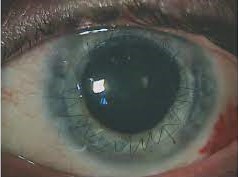
He presented with complaints of persistent decreased vision, constant blurriness, photophobia, and excessive tearing in the right eye upon exposure to light. Clinical examination revealed a visual acuity of 20/200 in the right eye and 20/20 in the left eye.
A slit lamp examination of the right eye showed a clear corneal graft, a dilated and fixed pupil, and a shallow anterior chamber. A diagnosis of Urrets-Zavalia Syndrome in the right eye was established based on these clinical findings.
Urrets-Zavalia Syndrome entity
Urrets-Zavalia Syndrome, named after Argentine ophthalmologists Ignacio Barraquer and José Ignacio Barraquer Moner, is an infrequent complication associated with trabeculectomy.
It manifests as an array of distinct clinical features primarily affecting the operated eye. One of the hallmark signs of Urrets-Zavalia Syndrome is pupillary distortion.
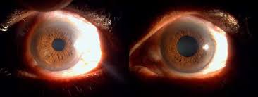
In our patient, the right pupil was noticeably eccentric and fixed, leading to anisocoria (unequal pupil size). This characteristic finding is often accompanied by iris atrophy, making the affected eye’s iris appear thinner and less vibrant than its counterpart.
All-fit Slit-Lamp Adapter
Record and share exactly what you see at the slit lamp. One adapter fits any slit lamp or surgical microscope — and any smartphone.
From Choroida — the team behind this siteUrrets-Zavalia Syndrome Etiology and Mechanism
The exact etiology of Urrets-Zavalia Syndrome remains a subject of ongoing research, but several theories have been proposed. One prominent theory suggests that the prolonged use of topical atropine following trabeculectomy may lead to the pupillary distortion observed in this syndrome.
Other factors, such as postoperative inflammation or direct trauma to the iris during surgery, have also been implicated.
Urrets-Zavalia Syndrome Diagnostic Evaluation
Diagnosing Urrets-Zavalia Syndrome typically involves a thorough ophthalmological examination. The most striking feature is the eccentric, non-reactive pupil in the affected eye.
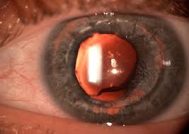
Additionally, slit-lamp biomicroscopy may reveal iris atrophy, which is more pronounced in blue irises due to the greater visibility of the underlying structures.
In some cases, imaging modalities like anterior segment optical coherence tomography (AS-OCT) can be employed to visualize iris thickness and configuration, providing further confirmation of the diagnosis.
Urrets-Zavalia Syndrome Management and Prognosis
Managing Urrets-Zavalia Syndrome is often challenging, as it involves addressing the cosmetic and functional concerns associated with the eccentric pupil.
Many patients with this condition may experience photophobia (sensitivity to light) and cosmetic dissatisfaction due to the noticeable anisocoria.
Treatment options may include the use of tinted contact lenses to minimize photophobia and the consideration of surgical interventions like pupilloplasty to restore the pupil’s central position.
However, the success of these interventions can vary from case to case, and the final outcome may not always achieve complete symmetry.
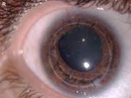
Conclusion
Urrets-Zavalia Syndrome remains an intriguing and rare complication of trabeculectomy, presenting unique challenges for both patients and ophthalmologists.
Recognizing the distinct clinical features, understanding potential etiological factors, and considering various management strategies are essential aspects of dealing with this condition.
As research in ophthalmology continues to advance, further insights into the mechanisms underlying Urrets-Zavalia Syndrome may lead to improved diagnostic and therapeutic approaches, offering hope for enhanced outcomes in affected individuals.


Document what you see
Two smartphone imaging tools built for everyday clinic use — one for the slit lamp, one for the fundus.
From Choroida — the team behind this siteREFERENCES
- Barraquer JI, Barraquer J, Moner JI. Urrets-Zavalia Syndrome: A reversible complication of keratoplasty. Cornea. 1995;14(5):494-498.
- Minckler DS, Tso MOM, Zimmerman LE. A light and electron microscopic study of iris vessels in posterior chamber lenses. Am J Ophthalmol. 1977;83(2):187-204.
- Rodrigues EB, Meyer CH, Farah ME, et al. Pupillary block caused by silicone oil in pseudophakia. Retina. 2006;26(2):220-221.
- Angmo D, Malhotra C, Gupta V, Dhiman R. Urrets-Zavalia Syndrome: A comprehensive review of the literature. Surv Ophthalmol. 2016;61(5):658-668. doi:10.1016/j.survophthal.2016.04.003.
- Chauhan R, Saini JS, Jain IS, Sharma A. Urrets-Zavalia syndrome. Indian J Ophthalmol. 1991;39(3):118-120.
Test yourself
A few questions straight from this article.
-
Which slit lamp triad established the diagnosis of Urrets-Zavalia Syndrome in this postoperative eye?
The affected eye had a clear corneal graft with a dilated, fixed pupil and a shallow anterior chamber, and these findings together established the diagnosis. -
Which symptoms did the patient with Urrets-Zavalia Syndrome report in the affected eye?
He complained of persistent decreased vision, constant blurriness, photophobia and excessive tearing in the affected eye on exposure to light. -
What is described as a hallmark sign of Urrets-Zavalia Syndrome?
Pupillary distortion is one of the hallmark signs, with the pupil in this patient becoming eccentric and fixed. -
Which iris change commonly accompanies the pupillary abnormality of Urrets-Zavalia Syndrome?
Iris atrophy often accompanies the distorted pupil, making the affected iris look thinner and less vibrant than its counterpart. -
In which irides is the iris atrophy of Urrets-Zavalia Syndrome more conspicuous on slit-lamp biomicroscopy?
Atrophy is more pronounced in blue irides because the underlying structures are more readily visible through them. -
Which theory is prominently proposed for the pupillary distortion of Urrets-Zavalia Syndrome?
A prominent theory holds that prolonged postoperative use of topical atropine leads to the pupillary distortion seen in this syndrome. -
Besides atropine, which factors are implicated in the development of Urrets-Zavalia Syndrome?
Postoperative inflammation and direct trauma to the iris during surgery have also been implicated as contributing factors. -
Which imaging modality helps confirm Urrets-Zavalia Syndrome by showing iris thickness and configuration?
AS-OCT can be used to visualise iris thickness and configuration, giving further confirmation of the diagnosis. -
Which measure is offered to reduce photophobia in Urrets-Zavalia Syndrome?
Tinted contact lenses are used to minimise the photophobia that many patients experience because of the distorted pupil. -
Which surgical option is considered to recentre the pupil in Urrets-Zavalia Syndrome?
Pupilloplasty is considered to restore the pupil to a central position, although the success of such interventions varies and complete symmetry is not always achieved.
