
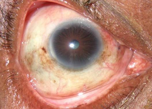
Leprosy (Hansen’s disease), caused by Mycobacterium leprae, remains an underappreciated cause of ocular morbidity in endemic regions despite the sharp global decline in overall case numbers since multidrug therapy became widely available.
Ocular involvement is common in longstanding, poorly treated disease and is a major contributor to disability in leprosy survivors, independent of the skin and nerve findings that usually dominate the clinical picture.
The organism has a predilection for cooler body surfaces, which is why the anterior segment of the eye — cornea, iris, and periocular skin — bears the brunt of ocular disease rather than the warmer posterior segment.
Nerve involvement compounds the problem, because facial and trigeminal nerve damage strips the eye of both its protective blink reflex and its corneal sensation at the same time.
Mechanisms of Ocular Damage
Direct bacillary invasion of ocular tissue produces lepromatous keratitis and iridocyclitis in multibacillary disease, while nerve damage from the immune response to the organism causes a separate, indirect set of problems: facial nerve (cranial nerve VII) involvement produces lagophthalmos, and trigeminal (cranial nerve V) involvement produces corneal anesthesia.
The combination is what makes leprosy-related corneal disease so severe, because neither protective mechanism (blink or pain) is available to warn the patient or protect the ocular surface.
Chronic low-grade inflammation from bacillary antigens can also produce a slowly progressive keratitis independent of any acute reactional episode, contributing to corneal opacification over years even in patients who are adherent to treatment.
Ocular and Periocular Findings
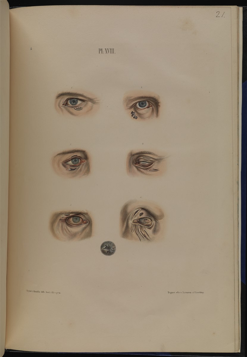
- Madarosis — loss of eyebrows and eyelashes, often an early and visible sign
- Lagophthalmos from facial nerve involvement
- Corneal hypoesthesia or anesthesia from trigeminal nerve involvement
- Punctate and diffuse superficial keratitis, sometimes progressing to corneal opacification
- Iris pearls — small, glistening nodules on the iris surface, relatively specific to lepromatous disease
- Chronic iridocyclitis, which can be low-grade and easily overlooked without a slit-lamp exam
- Cataract, often secondary to chronic uveitis
Because corneal sensation is frequently reduced, patients with leprosy-related keratitis often do not report the pain that would normally prompt an eye exam, which is a major reason ocular disease in leprosy tends to be diagnosed late.
All-fit Slit-Lamp Adapter
Record and share exactly what you see at the slit lamp. One adapter fits any slit lamp or surgical microscope — and any smartphone.
From Choroida — the team behind this siteLepra Reactions and the Eye
Type 1 (reversal) and type 2 (erythema nodosum leprosum) reactions are immune-mediated episodes that can flare during or after treatment and often worsen ocular inflammation acutely.
A patient with known leprosy who develops a sudden red, painful eye during a systemic reaction needs prompt ophthalmic assessment, because acute iridocyclitis during a reactional episode can progress quickly if undertreated, and a delay in recognizing this specific complication carries real risk to vision.
Systemic corticosteroids used to manage severe reactions also have a role in controlling the associated ocular inflammation, though the underlying antimicrobial treatment needs to continue in parallel.
Differential Diagnosis
- Herpes zoster ophthalmicus — vesicular rash, dermatomal distribution, acute onset
- Sarcoidosis — granulomatous uveitis, but without the peripheral neuropathy or skin lesions of leprosy
- Neurotrophic keratopathy from other causes (herpetic disease, diabetic neuropathy, prior surgery)
- Facial nerve palsy from other causes (Bell’s palsy, tumor, trauma) when madarosis and skin lesions are absent
A careful skin and peripheral nerve exam usually separates leprosy from these alternatives; thickened peripheral nerves and hypopigmented, anesthetic skin patches are clues that point specifically toward Hansen’s disease, and are worth checking for specifically in a patient from an endemic area presenting with otherwise unexplained ocular surface findings.
Management
Multidrug antimicrobial therapy — the WHO-recommended combination of rifampicin, dapsone, and clofazimine for multibacillary disease — treats the underlying infection but does not reverse established nerve damage or corneal scarring, which is why ocular protection has to run alongside systemic treatment rather than being deferred until after it.
Ocular protection needs to run in parallel with systemic treatment: aggressive lubrication for exposure and corneal anesthesia, taping or a moisture chamber at night for lagophthalmos, and prompt treatment of any secondary microbial keratitis given how easily an anesthetic, exposed cornea can become infected without the patient noticing.
Surgical options — lid-loading procedures for lagophthalmos, tarsorrhaphy in severe exposure, cataract extraction once inflammation is controlled — are used selectively once the systemic disease is under control, with the timing coordinated to avoid operating during an active reactional episode.
Regular ophthalmic screening is recommended for all patients with multibacillary leprosy, because ocular disease can progress silently given the sensory loss that so often accompanies it, meaning a patient’s own subjective comfort cannot be relied upon as a marker of how the eye is actually doing.
Public Health Significance
Leprosy-related blindness is largely preventable with early systemic treatment and routine ocular screening, though access to eye care in many endemic areas remains limited relative to the dermatologic and neurologic aspects of the disease that receive more attention.
Integrating eye exams into standard leprosy care pathways is one of the more cost-effective ways to reduce disability in affected populations, since the systemic diagnosis and treatment infrastructure is often already in place — it is the routine addition of an eye exam to that existing pathway that is most often the missing piece.


Document what you see
Two smartphone imaging tools built for everyday clinic use — one for the slit lamp, one for the fundus.
From Choroida — the team behind this siteReferences
- World Health Organization. Guidelines for the Diagnosis, Treatment and Prevention of Leprosy.
- Courtright P, Lewallen S. Ocular manifestations of leprosy. Community Eye Health Journal.
- Daniel E, Ffytche TJ, Kempen JH, et al. Incidence of ocular complications in patients with multibacillary leprosy. British Journal of Ophthalmology.
- Malik AN, Morris RJ, Ffytche TJ. The prevalence of ocular complications in leprosy patients. Eye.
- American Academy of Ophthalmology. Basic and Clinical Science Course, Section 8: External Disease and Cornea.
Test yourself
A few questions straight from this article.
-
Why does leprosy damage the anterior segment rather than the posterior segment of the eye?
That predilection is why cornea, iris and periocular skin bear the brunt of ocular disease while the warmer posterior segment is spared. -
Which nerve lesions produce lagophthalmos and corneal anesthesia in leprosy?
Losing both at once strips the eye of its protective blink and its corneal sensation, which is what makes the resulting corneal disease so severe. -
Which early, visible periocular sign occurs in leprosy?
Madarosis is often among the first outwardly visible signs, well before the deeper corneal and uveal changes become apparent. -
Which iris finding is relatively specific to lepromatous leprosy?
They accompany a chronic iridocyclitis that is often low-grade and easily missed without a deliberate slit-lamp examination. -
Why is leprosy-related keratitis so often diagnosed late?
Without the pain that normally prompts an eye examination, patients do not present until the corneal damage is already established. -
How should a sudden red, painful eye during a lepra reaction be handled?
Systemic corticosteroids used for severe type 1 or type 2 reactions also help the ocular inflammation, while antimicrobial treatment continues in parallel. -
Which findings point specifically toward Hansen's disease in a patient with ocular surface signs?
A careful skin and peripheral nerve examination separates leprosy from herpes zoster ophthalmicus, sarcoidosis and other causes of neurotrophic keratopathy. -
Which drug combination does the WHO recommend for multibacillary leprosy?
It treats the infection but reverses neither established nerve damage nor corneal scarring, so ocular protection must run alongside rather than after it. -
What overnight measure protects the cornea in leprosy-related lagophthalmos?
This sits alongside aggressive lubrication and prompt treatment of any secondary microbial keratitis, which an anesthetic exposed cornea acquires easily. -
Which patients with leprosy need regular ophthalmic screening?
Ocular disease progresses silently given the accompanying sensory loss, so a patient's own comfort cannot be relied on as a marker of eye health.
