
Case Study
A 48-year-old man presented with progressive visual disturbances, including difficulty seeing in low-light conditions and a gradual decline in peripheral vision.
He reported no significant systemic health issues, although his family history was notable for retinal diseases in his father.
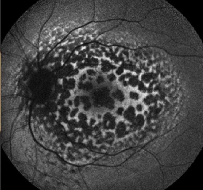
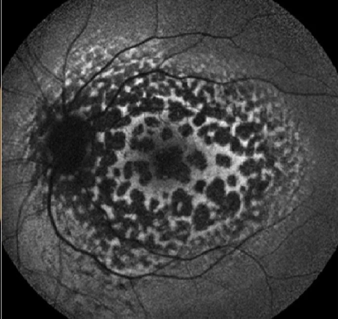
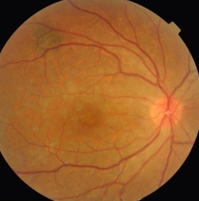
On examination, his visual acuity was 20/30 in both eyes. Fundoscopic evaluation revealed multiple, small, yellowish lesions scattered throughout the posterior pole, resembling the spots on a leopard.
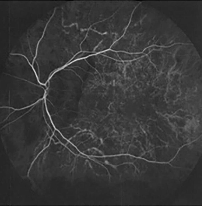
Optical coherence tomography (OCT) and fluorescein angiography confirmed pigmentary disturbances and areas of chorioretinal atrophy.
A diagnosis of Leopard Chorioretinopathy was established. Despite symptomatic management, his condition advanced, resulting in further visual impairment.
Disease Entity
Leopard Chorioretinopathy is a rare, chronic retinal disorder characterized by distinctive yellow or white lesions across the retina, resembling a leopard’s spots.
These lesions represent pigmentary alterations and areas of chorioretinal atrophy. Though the exact etiology remains uncertain, it is often associated with inherited retinal diseases or systemic syndromes such as choroideremia or Alport syndrome.
This condition predominantly affects the choroid and retinal pigment epithelium (RPE), leading to progressive vision loss.
Due to its rarity and overlapping features with other retinal dystrophies, Leopard Chorioretinopathy is frequently underdiagnosed or misclassified.
Pathophysiology
The pathogenesis of Leopard Chorioretinopathy involves degeneration of the RPE and choroid, resulting in characteristic chorioretinal lesions.
- RPE Degeneration: Loss of the RPE disrupts photoreceptor support, causing pigmentary changes and visual dysfunction.
- Choroidal Atrophy: Thinning and vascular compromise in the choroid contribute to ischemia and lesion development.
- Genetic Influence: Inherited mutations affecting retinal or choroidal integrity may underlie this condition.
While the precise molecular mechanisms remain unclear, studies suggest that oxidative stress and genetic predisposition play significant roles.
Epidemiology
Leopard Chorioretinopathy is an exceedingly rare condition, with limited cases documented in the literature.
- Age of Onset: Most cases manifest in mid-adulthood, though earlier presentations are possible.
- Gender Predilection: No significant gender predilection has been noted.
- Prevalence: It is primarily reported in individuals with underlying genetic or systemic conditions.
Clinical Features
Leopard Chorioretinopathy presents with a range of visual symptoms and distinct fundoscopic findings.
- Symptoms:
- Progressive decline in visual acuity.
- Difficulty seeing in dim light (nyctalopia).
- Peripheral visual field loss.
- Metamorphopsia in advanced cases.
- Fundoscopic Findings:
- Multiple yellow or white lesions are scattered across the posterior pole and mid-periphery.
- Pigmentary clumping or depigmentation.
- Choroidal thinning is visible in advanced stages.
- Imaging Features:
- OCT: Areas of RPE atrophy and photoreceptor loss.
- Fluorescein Angiography: Hyperfluorescent spots corresponding to choroidal lesions.
- Fundus Autofluorescence: Hypoautofluorescent areas denoting atrophic changes.
Diagnosis
The diagnosis of Leopard Chorioretinopathy requires a combination of clinical evaluation, imaging, and genetic testing.
- Clinical Examination:
- Comprehensive ocular examination, including fundoscopy, to identify characteristic lesions.
- Advanced Imaging:
- OCT to assess structural retinal changes.
- Fluorescein angiography and fundus autofluorescence for lesion mapping.
- Systemic Evaluation:
- Screening for associated systemic conditions, such as metabolic or genetic disorders.
- Genetic Testing:
- Identification of mutations linked to inherited retinal diseases, particularly in familial cases.
Differential Diagnosis
Leopard Chorioretinopathy shares clinical features with other retinal and choroidal conditions, necessitating careful differentiation.
- Central Areolar Choroidal Dystrophy (CACD): Central choroidal atrophy without diffuse lesions.
- Pattern Dystrophies: Retinal pigmentary changes localized to the macula.
- Choroideremia: Progressive choroidal atrophy with a genetic basis.
- Stargardt Disease: Fundus flecks and central vision loss, typically in younger patients.
Management
No definitive cure exists for Leopard Chorioretinopathy. Management focuses on symptom relief, visual rehabilitation, and addressing associated conditions.
- Ocular Interventions:
- Low-vision aids to enhance residual vision.
- Regular monitoring to assess progression.
- Systemic Therapy:
- Management of underlying systemic conditions or syndromes.
- Genetic counseling for hereditary cases.
- Experimental Therapies:
- Antioxidants and neuroprotective agents may slow progression, though evidence is limited.
Prognosis
The visual prognosis for Leopard Chorioretinopathy varies depending on disease severity and progression.
- Natural Course:
- Progressive vision loss is typical, with some patients retaining functional central vision.
- Factors Influencing Outcome:
- Early detection and management of systemic conditions.
- Access to visual rehabilitation resources.
Conclusion
Leopard Chorioretinopathy is a rare and enigmatic retinal condition marked by distinctive lesions and progressive visual impairment.
Its rarity, coupled with overlapping features with other retinal dystrophies, poses significant diagnostic and therapeutic challenges.
A multidisciplinary approach involving ophthalmologists, geneticists, and systemic disease specialists is crucial for optimal patient care.
Advances in genetic research and retinal imaging hold promise for improved understanding and management of this condition.


Document what you see
Two smartphone imaging tools built for everyday clinic use — one for the slit lamp, one for the fundus.
From Choroida — the team behind this siteReferences
- Shields JA, Shields CL. Retinal and choroidal manifestations of inherited dystrophies. Survey of Ophthalmology. 2015;60(5):425-432.
- Spaide RF. Autofluorescence imaging in retinal disease: Applications in Leopard Chorioretinopathy. American Journal of Ophthalmology. 2018;195:59-70.
- Braun DA, Hildebrandt F. Genetic determinants of retinal dystrophies with systemic associations. Journal of Clinical Investigation. 2017;127(8):2893-2902.
- Casselman B, Moosajee M. Insights into pattern dystrophies and Leopard Chorioretinopathy. Progress in Retinal and Eye Research. 2020;79:100854.
- Souied EH, Mrejen S. Imaging advances in rare retinal diseases. Retina Today. 2020;14(3):40-46.
Document what you see
Two smartphone imaging tools built for everyday clinic use — one for the slit lamp, one for the fundus.
From Choroida — the team behind this siteTest yourself
A few questions straight from this article.
-
Which fundus appearance gives Leopard Chorioretinopathy its name?
The condition is defined by distinctive yellow or white lesions across the retina resembling a leopard's spots, representing pigmentary change and chorioretinal atrophy. -
Which ocular layers are predominantly affected in Leopard Chorioretinopathy?
Leopard Chorioretinopathy predominantly affects the choroid and the retinal pigment epithelium, leading to progressive vision loss. -
Which systemic conditions are associated with Leopard Chorioretinopathy?
Although the exact etiology is uncertain, the condition is often associated with inherited retinal diseases or systemic syndromes such as choroideremia or Alport syndrome. -
At what stage of life do most cases of Leopard Chorioretinopathy present?
Most cases manifest in mid-adulthood, though earlier presentations are possible, and no significant gender predilection has been noted. -
Which symptom pattern is typical of Leopard Chorioretinopathy?
Patients report progressive decline in acuity, difficulty seeing in dim light, peripheral visual field loss, and metamorphopsia in advanced cases. -
What does fundus autofluorescence show in Leopard Chorioretinopathy?
Fundus autofluorescence reveals hypoautofluorescent areas that correspond to atrophic change, complementing OCT findings of RPE atrophy and photoreceptor loss. -
What does fluorescein angiography typically demonstrate in Leopard Chorioretinopathy?
Fluorescein angiography shows hyperfluorescent spots that correspond to the choroidal lesions, and is used alongside autofluorescence for lesion mapping. -
How does central areolar choroidal dystrophy differ from Leopard Chorioretinopathy?
Central areolar choroidal dystrophy produces central choroidal atrophy without the diffuse scattered lesions that characterise Leopard Chorioretinopathy. -
What is the mainstay of management in Leopard Chorioretinopathy?
No definitive cure exists; management centres on low-vision aids, regular monitoring for progression, treatment of associated systemic disease, and genetic counseling in hereditary cases. -
What is the expected natural course of Leopard Chorioretinopathy?
Progressive vision loss is typical, although some patients retain functional central vision; outcome depends on early detection and access to visual rehabilitation.
