
DISEASE
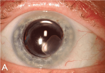
Ocular surface squamous neoplasia (OSSN) encompasses a wide and varied spectrum of diseases involving abnormal growth of dysplastic squamous epithelial cells on the surface of the eye.
- Conjunctival intraepithelial neoplasia (CIN) is non-invasive by definition; the basement membrane remains intact and the underlying substantia propria is spared. It is a slow-growing tumor that arises from a single mutated cell on the ocular surface. CIN is known by other names including Bowen’s disease, conjunctival squamous dysplasia, intraepithelial epithelioma, and epithelial dyskeratosis.
- Corneal epithelial dysmaturation, corneal epithelial dysplasia, and corneal intraepithelial neoplasia refer to neoplastic lesions of the cornea in which the conjunctival presence is minimal. The corneal epithelial islands or geographic epithelial granularity are the predominant clinical findings.
- Squamous cell carcinoma (SCC) describes a malignant lesion in which the dysplastic epithelial cells have penetrated the corneal basement membrane, gaining metastatic potential.
- Mucoepidermoid carcinoma represents a rare yet aggressive variant of SCC. It is, however, clinically indistinguishable from SCC and must be differentiated by histopathologic sampling.
According to epidemiology studies, the prevalence of Ocular surface squamous neoplasia (OSSN) is estimated to range from <0.2 cases/million/year (UK, 1996) to 35 cases/million/year (Uganda, 1992).
In several series, CIN has been reported to be the most common conjunctival neoplasia, whereas SCC has been found to be the most common conjunctival malignancy.
In the western hemisphere, OSSN afflicts mainly Caucasian men in their 60s to 70s who live close to the equator.
However, in Africa and certain parts of Asia, Ocular surface squamous neoplasia (OSSN) afflicts younger patients and tends to be more clinically aggressive.
A similar pattern has been observed in patients with the human immunodeficiency virus (HIV) and xeroderma pigmentosum.
The clinical presentation of Ocular surface squamous neoplasia (OSSN) is variable, making diagnosis sometimes difficult. Typically, patients present with a gelatinous or plaque-like interpalpebral conjunctival gray or white lesion.
Approximately 95% of CIN lesions occur at the limbus, where the most actively mitotic cells reside. The lesion may be flat or elevated and may be associated with feeder vessels.
Fluorescein, lissamine green, or Rose Bengal are often used to highlight the lesion. Abnormal or dysplastic epithelium has a diffuse, granular appearance, differentiating it from normal epithelium.
MANAGEMENT
General treatment:
The onset of the lesion, whether the lesions represent a recurrence, and prior treatments dictate tumor management.
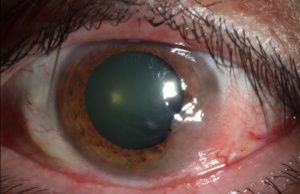
Major breakthroughs in the diagnosis and clinical approach of this disease have occurred within the past 2 to 3 decades. There has been an increasing trend toward employing medical therapy over surgery.
Medical therapy:
The use of topical chemotherapeutic agents, including Interferon-α2b, mitomycin C, and 5- 5-fluorouracil, has the advantage of treating the entire ocular surface and avoiding surgical complications such as positive margins, scarring, and limbal stem cell deficiency.
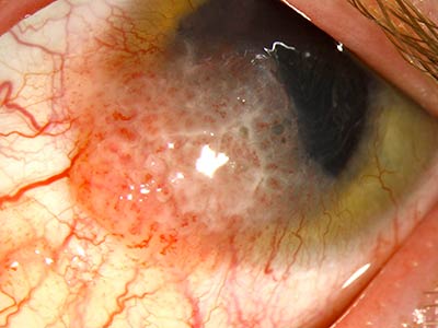
- Interferon-α2b (IFNα2b) is a cytokine produced by immune cells to combat microbes and viruses. Its mechanism of action is thought to be related to its antiproliferative, cytotoxic, antiviral, and antigenic properties. It may be injected subconjunctivally or used topically as an eye drop. Subconjunctival injections, concentrated at 3 million IU/0.5 mL, are administered weekly until resolution of the lesion. The concentration for the topical eye drop is typically 1 million IU/mL. Topical treatment is administered 4 times daily until 1-2 months after resolution of the lesion, which takes 4 months on average. The efficacy rate after topical IFNα2b ranges from 80% to 100%. Unfortunately, although well tolerated, topical IFNα2b is only available through specialized compounding pharmacies, requires refrigeration, and is costly.
- 5- fluorouracil (5-FU) blocks DNA synthesis by acting as a pyrimidine analog after incorporation into RNA. Its efficacy rate has been reported to be 100% after one to five cycles (1 month on and 3 months off), with a recurrence rate of up to 20%. Another regimen that causes less inflammation to the ocular surface involves cycling the medication 4 times daily for 4 to 7 days followed by a 3-week holiday. Treatment with 5-FU is less costly than MMC and IFNa2b and also has a favorable side effect profile compared with MMC. Disadvantages include mild ocular irritation and occasional conjunctivitis. Topical corticosteroids and preservative-free artificial tears may be co-administered to reduce these side effects.
- Topical mitomycin C (MMC) has proven to be an efficacious treatment of Ocular surface squamous neoplasia (OSSN). Mitomycin C is an antimetabolite that alkylates DNA and disrupts the production of RNA. Studies have reported its efficacy rate to range from 80% to 100%. MMC comes in either 0.02% or 0.04%. The lower concentration is usually prescribed continuously for a month; whereas, the higher concentration may be used for a week followed by 2 to 3 weeks of treatment. It must be refrigerated. While clinical resolution and recurrence rates are similar to IFN, the time to resolution is typically shorter with MMC. The disadvantages of MMC include ocular pain, possible limbal stem cell loss, and other ocular surface toxicity. Punctal plug occlusion is advised to decrease the risk of punctual stenosis.
- Reports have shown that both topical and subconjunctival injections of anti-vascular endothelial growth factor (VEGF) are efficacious in treating OSSN. One case series reported improvement in the size and vascularity of conjunctival Ocular surface squamous neoplasia (OSSN) after 3 months of treatment with subconjunctival injections of bevacizumab with little effect on cornea involvement. Another small case series reported a significant reduction in the size of the lesion with the use of topical bevacizumab in 10 eyes after 5 to 14 weeks.
Surgery:
A no-touch technique is used during the excision of Ocular surface squamous neoplasia (OSSN) lesions. CIN and SCC lesions involving the limbus should be excised with at least a 3-4mm uninvolved conjunctival margin.
A large conjunctival margin is important since seemingly uninvolved tissue clinically may still contain dysplastic cells.
Absolute alcohol is then applied to the cornea to loosen the epithelium from the basement membrane and then rinsed off with copious irrigation after 30 to 40 seconds.
All involved corneal epithelium and any associated corneal pannus are then scraped off with a Beaver blade or surgical sponge after the instillation of absolute alcohol to loosen the cells.
It is very important that the underlying Bowman’s layer is not violated during this process. Using a double or triple rapid freeze-thaw technique, cryotherapy is applied to the conjunctival edges, involved limbal zone, and the bare scleral bed to kill any remaining dysplastic cells.
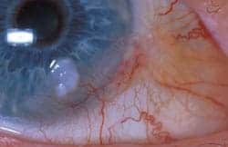
Cryotherapy is very important since it effectively extends the surgical margins. If the margins return positive or there are any concerns for residual disease, topical chemotherapy may be used after excision.
Both epithelial dysmaturation and corneal epithelial dysplasia are considered benign lesions and are treated with corneal scraping and wide conjunctival limbal margins if involved.
In cases of SCC, a thin lamellar scleral flap after the removal of the conjunctival lesion is advised followed by the application of absolute alcohol to the scleral bed. The area of surgical resection may be left open or closed with amniotic membrane graft.
In cases of local invasion into the eye or orbit, enucleation and exteneration may be done in collaboration with an oculoplastics surgeon.
Sentinel node biopsy may also be appropriate to stage the disease. Radiation may be considered as adjunctive therapy in certain cases that have been recalcitrant to other modalities of treatment.


Document what you see
Two smartphone imaging tools built for everyday clinic use — one for the slit lamp, one for the fundus.
From Choroida — the team behind this siteAll-fit Slit-Lamp Adapter
Record and share exactly what you see at the slit lamp. One adapter fits any slit lamp or surgical microscope — and any smartphone.
From Choroida — the team behind this siteREFERENCES
- Krachmer JH, Mannis MJ, Holland EJ. Cornea: Fundamentals, diagnosis and management. 2005.
- Reidy JJ, Bouchard CS, Florakis GJ, et al. Basic and Clinical Science Course, 2011-2012. 2011:226–233.
- Ateenyi-Agaba C. Conjunctival squamous-cell carcinoma associated with HIV infection in Kampala, Uganda. The Lancet 1995;345:695–696.
- Newton R, Reeves G, Beral V, et al. Effect of ambient solar ultraviolet radiation on incidence of squamous-cell carcinoma of the eye. The Lancet 1996;347:1450–1451.
- Karp CL, Scott IU, Chang TS, Pflugfelder SC. Conjunctival Intraepithelial Neoplasia: A Possible Marker for Human Immunodeficiency Virus Infection? Arch Ophthalmol 1996;114:257–261.
Test yourself
A few questions straight from this article.
-
What does ocular surface squamous neoplasia describe?
OSSN covers a wide spectrum of disease defined by abnormal growth of dysplastic squamous epithelial cells on the surface of the eye. -
What makes conjunctival intraepithelial neoplasia non-invasive by definition?
CIN is non-invasive by definition: the basement membrane remains intact and the underlying substantia propria is spared. It is a slow-growing tumour arising from a single mutated cell. -
What defines squamous cell carcinoma within the ocular surface squamous neoplasia spectrum?
SCC is a malignant lesion in which the dysplastic epithelial cells have penetrated the basement membrane, thereby gaining metastatic potential. -
How is mucoepidermoid carcinoma of the ocular surface distinguished from squamous cell carcinoma?
Mucoepidermoid carcinoma is a rare but aggressive variant of SCC that is clinically indistinguishable from it, so histopathologic sampling is required to tell them apart. -
Approximately what proportion of conjunctival intraepithelial neoplasia lesions arise at the limbus?
About 95% of CIN lesions occur at the limbus, the site where the most actively mitotic cells of the ocular surface reside. -
What is the typical clinical appearance of ocular surface squamous neoplasia?
Patients typically present with a gelatinous or plaque-like interpalpebral conjunctival grey or white lesion, flat or elevated, sometimes with feeder vessels. -
Which dyes are used to highlight an ocular surface squamous neoplasia lesion at the slit lamp?
Fluorescein, lissamine green or Rose Bengal are often used to highlight the lesion; dysplastic epithelium takes on a diffuse, granular appearance unlike normal epithelium. -
What advantage do topical chemotherapeutic agents offer over surgical excision in OSSN?
Topical agents such as interferon-alpha2b, mitomycin C and 5-fluorouracil treat the entire ocular surface and avoid surgical complications including positive margins, scarring and limbal stem cell deficiency. -
What conjunctival margin is advised when excising limbal CIN or squamous cell carcinoma?
At least a 3-4 mm uninvolved conjunctival margin is recommended, because tissue that looks uninvolved clinically may still contain dysplastic cells. -
Why is cryotherapy applied during excision of ocular surface squamous neoplasia?
Cryotherapy is applied by double or triple rapid freeze-thaw to the conjunctival edges, involved limbal zone and bare scleral bed to kill remaining dysplastic cells, effectively extending the surgical margins.

Writes and reviews the clinical articles on Educate. Every post is checked against peer-reviewed sources before it goes live.


