
In 2018, an algorithm became the first AI diagnostic system in any field of medicine to receive FDA authorisation to make a diagnosis without a physician reviewing the image.
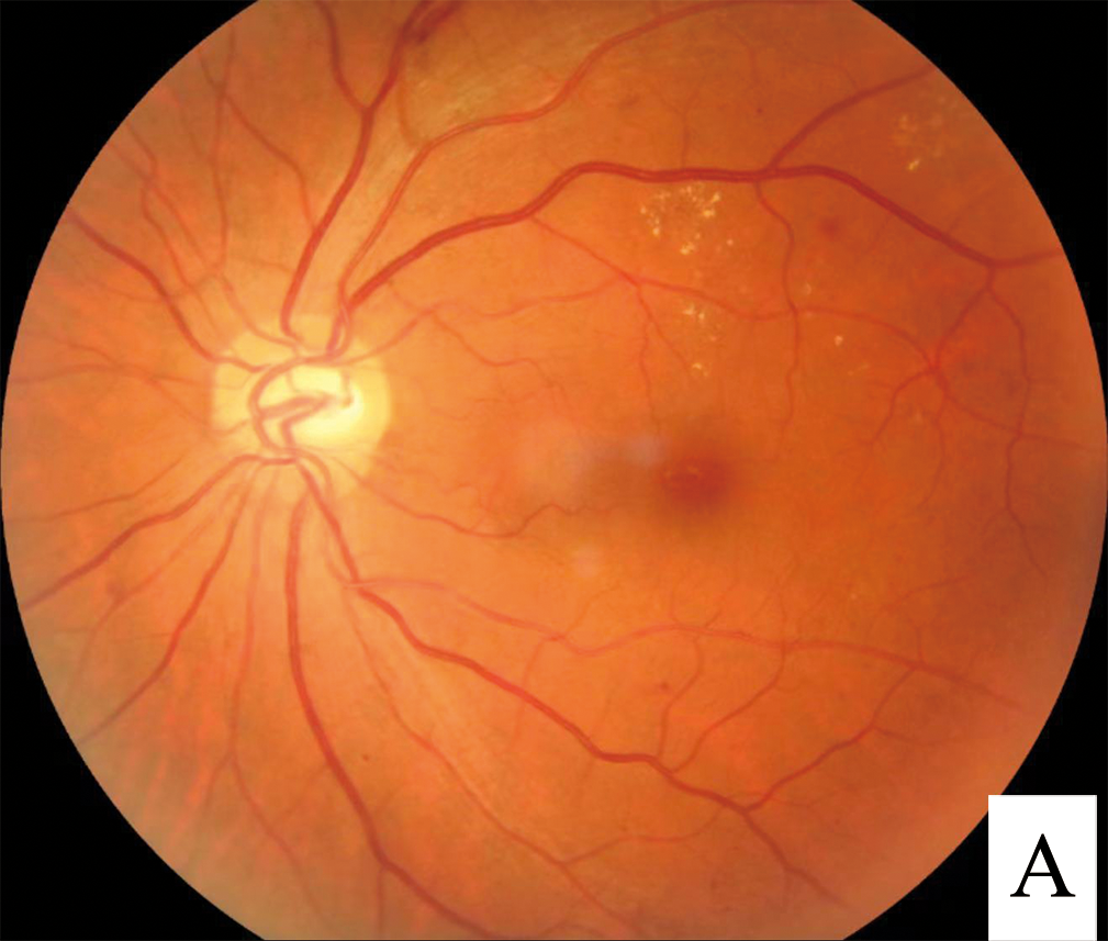
The disease it diagnosed was diabetic retinopathy, and the image was an ordinary fundus photograph.
AI-based diabetic retinopathy screening is now deployed at scale in primary care and community settings that have never had access to an ophthalmologist.
The clinical case for it is almost entirely about a mismatch: the number of people with diabetes is enormous, and the number of eye specialists able to screen them all, every year, is not.
Understanding what these systems can and cannot do is now a basic requirement for anyone practising in diabetic eye care.
What Is AI Diabetic Retinopathy Screening?
AI diabetic retinopathy screening uses deep learning algorithms, typically convolutional neural networks, to analyse fundus photographs and automatically detect referable diabetic retinopathy without a human grader in the loop.
Systems in current use fall into two broad categories:
- Autonomous systems, authorised to issue a screening result directly to the patient or primary care provider without human over-read
- Assistive systems, which flag or pre-grade images for a human reader, aiming to reduce grading workload rather than replace it entirely
The regulatory distinction between these two categories matters enormously – autonomous systems carry a different liability and validation burden than assistive tools sitting alongside a human grader.
The Screening Problem This Solves
- The number of people with diabetes worldwide has risen steeply, and annual retinal screening is recommended for essentially all of them
- Trained human graders and ophthalmologists cannot scale to meet that volume in many health systems, particularly in low-resource and rural settings
- Manual grading is also subject to inter-grader variability, meaning even fully staffed programmes are not perfectly consistent
- Non-attendance at screening remains one of the largest single drivers of preventable diabetic vision loss, independent of grading accuracy
The core value proposition is capacity, not just accuracy – these tools exist primarily to screen people who would otherwise not be screened at all.
Fundus Explorer Pro
Photograph the retinal findings described here with the phone already in your pocket — 22 D optics and built-in illumination in one handheld unit.
From Choroida — the team behind this siteHow the Technology Works
- Deep convolutional neural networks are trained on large datasets of fundus images labelled by expert human graders against a reference standard, typically the International Clinical Diabetic Retinopathy severity scale
- The network learns to detect the lesions that define disease severity – microaneurysms, dot and blot haemorrhages, hard exudates, cotton wool spots and neovascularisation
- Output is typically a binary or graded classification – referable versus non-referable disease, sometimes with severity staging
- Image quality assessment is built into most systems, since poor-quality images that cannot be reliably graded need to be flagged rather than misclassified
What the Model Actually Sees

A gradable colour fundus photograph, of the kind captured by tabletop or handheld retinal cameras, is the raw input for these algorithms – the same image type a human grader would review, but processed in seconds rather than minutes.
Clinical Evidence
- IDx-DR became the first autonomous AI diagnostic system authorised by the FDA for any field of medicine, based on a prospective trial demonstrating sensitivity and specificity meeting pre-specified thresholds for detecting more-than-mild diabetic retinopathy
- Multiple subsequent systems, including tools developed and validated across large screening populations in the UK, India and elsewhere, have shown comparable performance to human graders on external validation datasets
- Real-world deployment studies have generally shown good sensitivity for referable disease but performance can vary by population, camera type and image quality compared with the controlled conditions of pivotal trials
- Cost-effectiveness analyses generally favour AI-assisted screening for populations with capacity constraints, though the calculation depends heavily on local staffing costs and screening uptake
The gap between pivotal-trial performance and real-world performance is the recurring theme in this literature – external validation on a program’s own population and camera is not optional before deployment.
Integration Into Screening Pathways
Typical Autonomous Workflow
- Fundus photographs are captured, often by a technician or trained non-specialist in a primary care or community setting
- The algorithm grades image quality and disease status in real time or near real time
- Patients graded as non-referable are recalled per the standard screening interval; referable results are sent for prompt ophthalmic assessment
- Ungradable images are routed to human review or a repeat photograph
Assistive Workflow
- The algorithm pre-grades or triages images before a human grader review, aiming to prioritise likely-referable cases and reduce grader workload on clearly normal images
- A human grader remains the final decision-maker in every case
Ungradable-image handling is one of the most practically important design decisions in any of these systems – a program that quietly discards or mishandles ungradable images loses exactly the patients most in need of a careful look.
Limitations and Safety Considerations
- Algorithms are trained and validated for diabetic retinopathy specifically, and are not general-purpose fundus interpretation tools – they can miss unrelated pathology such as glaucomatous disc changes or macular degeneration unless separately trained and validated for those conditions
- Performance can degrade on populations, ethnicities or camera types under-represented in training data, which is why local validation before deployment is now widely recommended
- Image quality remains a major failure point – poor pupil dilation, media opacity and patient movement all reduce gradability
- A clear escalation pathway and clinical governance structure is required for referable results, ungradable images and any diagnostic disagreement
- Regulatory approval in one jurisdiction or for one algorithm version does not automatically extend to another population, camera, or software update
The single-disease scope of these tools is the point most likely to be misunderstood by non-specialist users – a normal diabetic retinopathy screening result says nothing about the optic nerve or macula in isolation.
Prognosis for the Field
AI screening is moving from novelty to standard infrastructure in diabetic eye care.
- Multiple national screening programmes have already incorporated AI grading, either autonomously or as an assistive first pass
- Expansion toward multi-disease screening – simultaneous detection of glaucomatous and age-related macular changes from the same image – is an active area of development
- Continued external validation, post-deployment monitoring and clear accountability structures remain the main barriers to broader trust and adoption, more than the underlying algorithmic accuracy
- The realistic long-term role is not replacing ophthalmologists but extending screening reach to populations who currently receive none at all
The technology has already answered the accuracy question convincingly – what remains is largely a health-systems question of deployment, governance and equitable access.


Document what you see
Two smartphone imaging tools built for everyday clinic use — one for the slit lamp, one for the fundus.
From Choroida — the team behind this siteReferences
- Abramoff MD, Lavin PT, Birch M, et al. Pivotal trial of an autonomous AI-based diagnostic system for detection of diabetic retinopathy. npj Digital Medicine. 2018.
- Gulshan V, Peng L, Coram M, et al. Development and validation of a deep learning algorithm for detection of diabetic retinopathy in retinal fundus photographs. JAMA. 2016.
- Ting DSW, Cheung CY, Lim G, et al. Development and validation of a deep learning system for diabetic retinopathy and related eye diseases. JAMA. 2017.
- Artificial Intelligence in Diabetic Retinopathy Screening. EyeWiki, American Academy of Ophthalmology.
- Diabetic Retinopathy Screening. StatPearls, NCBI Bookshelf.
Test yourself
A few questions straight from this article.
-
Which network architecture underlies current AI diabetic retinopathy screening systems?
AI diabetic retinopathy screening uses deep learning algorithms, typically convolutional neural networks, to analyse fundus photographs for referable disease. -
What distinguishes an assistive from an autonomous AI screening system for diabetic retinopathy?
Assistive systems pre-grade or triage images to reduce grader workload, but a human grader remains the final decision-maker in every case. -
What made IDx-DR notable when it received FDA authorisation?
IDx-DR was the first autonomous AI diagnostic system authorised by the FDA in any field of medicine, based on a prospective trial detecting more-than-mild diabetic retinopathy. -
Against which reference standard are training images for these algorithms typically labelled?
Networks are trained on large datasets of fundus images labelled by expert human graders against a reference standard, typically the International Clinical Diabetic Retinopathy severity scale. -
Which set of lesions are diabetic retinopathy screening algorithms trained to detect?
The network learns the lesions defining severity: microaneurysms, dot and blot haemorrhages, hard exudates, cotton wool spots and neovascularisation. -
How should an autonomous screening pathway handle an image the algorithm cannot grade?
Image quality assessment is built in so ungradable images are flagged rather than misclassified, then routed to human review or a repeat photograph. -
What is the core value proposition of AI diabetic retinopathy screening?
The value is capacity rather than accuracy alone: these tools exist primarily to screen people who would otherwise receive no screening at all. -
A normal AI diabetic retinopathy screening result leaves which question unanswered?
These algorithms are validated for diabetic retinopathy specifically and can miss unrelated pathology such as glaucomatous disc changes or macular degeneration. -
Why is local validation recommended before deploying an AI screening algorithm?
Performance can degrade on populations, ethnicities or camera types under-represented in the training data, so external validation on the programme's own population is not optional. -
What now represents the main barrier to wider adoption of AI retinopathy screening?
Continued external validation, post-deployment monitoring and clear accountability remain the main barriers to broader trust and adoption, more than algorithmic accuracy.

Writes and reviews the clinical articles on Educate. Every post is checked against peer-reviewed sources before it goes live.


