
CASE REPORT
A 71-year-old woman with diabetes and hypertension presented with complaints of incomplete recovery of vision post right eye cataract surgery performed 2 months prior to presentation.

On examination, the best-corrected visual acuity in the right eye was 20/40 p, N6, and the left eye was 20/50, N6.
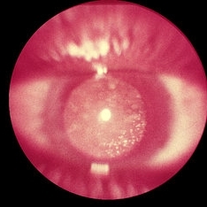
Her right eye examination showed prolapse of the vitreous in the anterior chamber with incarceration in the inner lip of the superotemporal limbal incisional scar causing peaking of the pupil along with temporal decentration of the intraocular lens.
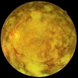
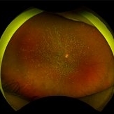
There were multiple white dots with pigments seen in this vitreous framework. On posterior segment examination, dense asteroid hyalosis (AH) was observed with a hazy view of the underlying disc and retina.
DISEASE
Asteroid hyalosis (AH) is a common clinical entity in which calcium-lipid complexes are suspended throughout the collagen fibrils of the vitreous.
Benson, in 1894, was the first to describe accurately and to differentiate AH from synchysis scintillans. Because the vitreous particles resembled “stars on a clear night” he termed the condition asteroid hyalitis, but Luxenberg and Sime later suggested the term “asteroid hyalosis” in view of the absence of inflammatory changes.
Clinically, the AH granules move with the movement of the eye and do not gravitate downwards like synchysis scintillans.
Histologically, asteroid bodies are rounded structures that stain positively with alcian blue and positively with stains for neutral fats, phospholipids, and calcium. The bodies stain metachromatically and exhibit birefringence.
Occasionally, asteroid bodies will be surrounded by a foreign body giant cell, but the condition is not generally associated with vitreous inflammation.
MANAGEMENT
Asteroid Hyalosis rarely causes visual disturbances, and surgical removal is only rarely required. There have been a few reports of AH in patients with RP.
In some cases, even a standard three-port vitrectomy is needed to remove the vitreous opacity because of the progression of the vitreous opacity and decreased vision.
Would you have interest in taking retina images by smartphone?
Fundus photography is superior to fundus analysis as it enables intraocular pathologies to be photo-captured and encrypted information to be shared with colleagues and patients.
Recent technologies allow smartphone-based attachments and integrated lens adaptors to transform the smartphone into a portable fundus camera and Retinal imaging by smartphone.
RETINAL IMAGING BY YOUR SMARTPHONE
REFERENCES
- Ophthalmology, A.A.o., Section 12: Retina and Vitreous. Basic and Clinical Science Course. 2014-2015: American Academy of Ophthalmology. p. 311-312.
- Yazar, Z., et al., Asteroid hyalosis. Eur J Ophthalmol, 2001. 11(1): p. 57-61.
- Shields, C.L., et al., Vitreous asteroid hyalosis prolapse into the anterior chamber simulating iris metastasis. Middle East Afr J Ophthalmol, 2012. 19(3): p. 346-8.
- Mitchell, P., M.Y. Wang, and J.J. Wang, Asteroid hyalosis in an older population: the Blue Mountains Eye Study. Ophthalmic epidemiology, 2003.
- Moss, S.E., R. Klein, and B.E.K. Klein, Asteroid hyalosis in a population: The Beaver Dam Eye Study. American Journal of Ophthalmology, 2001. 132(1): p. 70-75.
