
For decades, treating retinopathy of prematurity meant one thing: laser to the peripheral retina, delivered under strict time pressure once a threshold was crossed.
Then a single trial – BEAT-ROP – showed that an anti-VEGF injection could outperform laser in the most aggressive, posterior form of the disease.
Treating retinopathy of prematurity (ROP) is no longer a one-technique field, and the choice between laser and anti-VEGF now shapes the entire follow-up plan for these infants.
Both options work by the same underlying logic – interrupting VEGF-driven neovascularisation – but they diverge sharply in what happens afterward.
Getting that choice right, in an infant whose retina is still developing, carries consequences that last a lifetime.
What Is Retinopathy of Prematurity, and When Is It Treated?
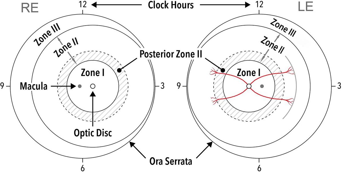
Retinopathy of prematurity is abnormal, VEGF-driven retinal vascularisation occurring in preterm infants whose retinal blood vessels had not finished developing at birth.
Treatment decisions rest on internationally standardised classification:
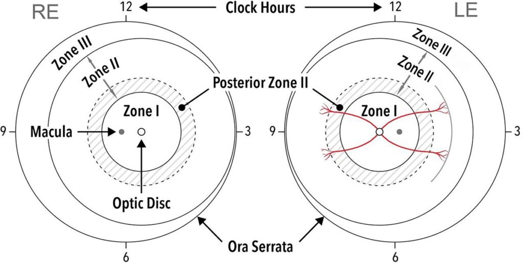
- Zone – how far posteriorly the avascular retina extends, with zone I disease being the most posterior and highest risk
- Stage – the severity of the vascular-avascular junction, from a flat demarcation line (stage 1) through to retinal detachment (stage 4-5)
- Plus disease – dilated, tortuous posterior vessels indicating high disease activity
Type 1 ROP – the accepted treatment threshold – is defined as zone I disease of any stage with plus disease, zone I stage 3 without plus disease, or zone II stage 2 or 3 with plus disease.
Why Treatment Is Time-Critical
- Type 1 ROP carries a substantial risk of progression to retinal detachment if untreated
- The Early Treatment for ROP study established that treating at the type 1 threshold, rather than waiting for older threshold criteria, significantly reduces unfavourable outcomes
- Screening schedules are calculated from gestational age and birth weight specifically so that no infant crosses the treatment threshold between examinations
- Once type 1 disease is identified, treatment is generally delivered within 48 to 72 hours
The screening programme and the treatment threshold are really one system – treatment only works this well because the screening interval is short enough to catch disease as it crosses the line.
Fundus Explorer Pro
Photograph the retinal findings described here with the phone already in your pocket — 22 D optics and built-in illumination in one handheld unit.
From Choroida — the team behind this siteLaser Photocoagulation
Technique
- Ablation of the avascular peripheral retina anterior to the vascular-avascular junction, using indirect ophthalmoscope-delivered diode laser
- Performed under general or sedation-based anaesthesia given the extent of treatment required in a small eye
- Confluent burns are placed across the entire avascular zone in one or, occasionally, staged sessions
Outcomes and Limitations
- Long track record and durable, well-characterised structural outcomes
- Causes permanent destruction of peripheral retina, with resulting peripheral visual field loss
- Associated with myopia, sometimes high myopia, later in childhood
- Technically difficult in zone I disease and in eyes with significant plus disease and hazy media
Laser remains the more predictable, better-understood option, which is why it has not simply been replaced despite anti-VEGF’s advantages in specific situations.
Anti-VEGF Therapy
Technique
- Intravitreal bevacizumab, and increasingly ranibizumab, injected using a scaled-down version of adult intravitreal injection technique
- A single injection can address the entire avascular retina, without a need to reach every point with a laser probe
Evidence and Rationale
- The BEAT-ROP trial showed intravitreal bevacizumab was superior to laser for zone I, stage 3+ disease specifically, with lower recurrence in this posterior subgroup
- Anti-VEGF avoids immediate destruction of peripheral retina, theoretically preserving future peripheral visual field and reducing induced myopia
- Continued peripheral vascularisation can occur after anti-VEGF treatment, unlike after laser, which is felt to explain some of the field and refractive advantages seen
Key Limitations
- Recurrence after anti-VEGF can occur later than after laser – sometimes many weeks post-injection – requiring a longer and less predictable follow-up window
- Long-term systemic safety data in this population remain more limited than the extensive laser experience, an important discussion point with families
- Access to appropriate follow-up imaging and examination over an extended period is essential, and not available in every setting
The extended, unpredictable recurrence window is the single biggest practical argument against anti-VEGF as a default choice – it demands a follow-up commitment that laser does not.
Choosing Between Laser and Anti-VEGF
- Zone I disease, particularly aggressive posterior ROP, favours anti-VEGF based on BEAT-ROP evidence
- Zone II disease is commonly treated with laser, which remains highly effective and avoids the extended recurrence-monitoring burden
- Media opacity or difficult visualisation for laser favours the injection route
- Reliability of long-term follow-up access is a major practical factor – anti-VEGF is a poor choice where sustained monitoring cannot be guaranteed
- Combination approaches – anti-VEGF followed by planned laser to the persistently avascular retina – are used in some centres to capture benefits of both
This decision increasingly sits with a specialist retina service rather than being a fixed protocol, precisely because the right answer depends on zone, media clarity and the realities of local follow-up capacity.
Complications of Treatment
- Laser – peripheral field loss, induced myopia, rare choroidal effusion or cataract from inadvertent lens or ciliary body treatment
- Anti-VEGF – late recurrence requiring vigilant extended follow-up, theoretical systemic VEGF suppression effects in a developing infant, and the same rare intravitreal injection risks seen in adults
- Both – progression to retinal detachment despite treatment in the most severe or rapidly progressive cases, which then requires vitreoretinal surgery
Systemic VEGF is essential for normal organ development in a premature infant, which is precisely why the safety question around anti-VEGF in this population is taken so seriously and remains an active area of research.
Prognosis
Outcomes have improved dramatically since routine screening and treatment protocols were established.
- Type 1 ROP treated promptly with either modality achieves favourable structural outcomes in the large majority of eyes
- Zone I and aggressive posterior disease carry a higher risk of unfavourable outcome despite treatment, reflecting the severity of the underlying disease rather than treatment failure
- Long-term follow-up into childhood is required regardless of treatment choice, given the risks of myopia, strabismus, amblyopia and late retinal detachment
- Progression to retinal detachment despite treatment carries a guarded visual prognosis and requires urgent vitreoretinal surgical referral
The direction of travel in this field is toward less destructive treatment – the question is no longer only whether ROP can be treated, but which treatment leaves a child with the best possible visual field and refractive outcome decades later.


Document what you see
Two smartphone imaging tools built for everyday clinic use — one for the slit lamp, one for the fundus.
From Choroida — the team behind this siteReferences
- Mintz-Hittner HA, Kennedy KA, Chuang AZ, BEAT-ROP Cooperative Group. Efficacy of intravitreal bevacizumab for stage 3+ retinopathy of prematurity. New England Journal of Medicine. 2011.
- Early Treatment for Retinopathy of Prematurity Cooperative Group. Revised indications for treatment of ROP. Archives of Ophthalmology. 2003.
- International Committee for the Classification of Retinopathy of Prematurity. The International Classification of ROP revisited. Archives of Ophthalmology. 2005.
- Retinopathy of Prematurity Treatment. EyeWiki, American Academy of Ophthalmology.
- Retinopathy of Prematurity. StatPearls, NCBI Bookshelf.
Test yourself
A few questions straight from this article.
-
Which finding meets the type 1 threshold for treating retinopathy of prematurity?
Type 1 ROP is zone I disease of any stage with plus disease, zone I stage 3 without plus disease, or zone II stage 2 or 3 with plus disease. -
What defines plus disease in retinopathy of prematurity?
Plus disease means dilated, tortuous posterior vessels, and it is the sign that indicates high disease activity within the zone and stage classification. -
Once type 1 retinopathy of prematurity is identified, within what window is treatment generally delivered?
Type 1 disease carries a substantial risk of progression to retinal detachment, so treatment is generally delivered within 48 to 72 hours of identification. -
What did the Early Treatment for ROP study establish about when to intervene?
ETROP showed that treating at the type 1 threshold, rather than waiting for older threshold criteria, significantly reduces unfavourable outcomes. -
In which subgroup did BEAT-ROP show intravitreal bevacizumab superior to laser?
BEAT-ROP showed bevacizumab was superior to laser specifically for zone I stage 3 plus disease, with lower recurrence in this posterior subgroup. -
Which retina does laser photocoagulation ablate when treating retinopathy of prematurity?
Laser ablates the avascular peripheral retina anterior to the vascular-avascular junction, delivered as confluent burns through an indirect ophthalmoscope. -
Which refractive outcome is laser treatment for retinopathy of prematurity associated with later in childhood?
Laser causes permanent peripheral retinal destruction and is associated with myopia, sometimes high myopia, later in childhood. -
What is the main practical argument against choosing anti-VEGF as the default ROP treatment?
Recurrence after anti-VEGF can occur many weeks post-injection, later than after laser, demanding a longer and less predictable follow-up commitment. -
Why is the systemic safety of anti-VEGF taken so seriously in premature infants?
Systemic VEGF is essential for normal organ development in a premature infant, which is why systemic suppression remains an active area of research. -
Why does follow-up into childhood remain necessary after any ROP treatment?
Long-term follow-up is required regardless of treatment choice, given the risks of myopia, strabismus, amblyopia and late retinal detachment.

Writes and reviews the clinical articles on Educate. Every post is checked against peer-reviewed sources before it goes live.


