Distichiasis vs trichiasis is a common clinical question because both conditions produce lashes that rub the cornea.
However, the lashes arise from different places, and that origin guides treatment. Therefore, careful slit lamp examination comes before any epilation or surgery.
Defining Distichiasis vs Trichiasis
Distichiasis is an extra row of lashes emerging from or near the meibomian gland orifices (see distichiasis). Trichiasis, however, describes normal lashes from normal follicles that grow toward the globe (see trichiasis), and the lid margin itself may sit in a normal position in both conditions.
Where the Extra Lashes Come From
Meibomian gland tissue undergoes metaplasia into pilosebaceous units in distichiasis, and as a result the follicles sit within the tarsus of the posterior lamella. The lashes are often finer, shorter and paler than the normal row.
All-fit Slit-Lamp Adapter
Record and share exactly what you see at the slit lamp. One adapter fits any slit lamp or surgical microscope — and any smartphone.
From Choroida — the team behind this siteHow Normal Lashes Become Misdirected
Trichiatic lashes arise from the anterior lamella, where follicles normally live. However, inflammation or scarring around the follicle changes the direction of growth, and then a few lashes, or a whole segment, turn back against the cornea.
Causes of Distichiasis vs Trichiasis
Congenital distichiasis is often autosomal dominant and links to FOXC2 mutations (see lymphedema distichiasis). Acquired distichiasis follows severe posterior lamellar inflammation, for example Stevens-Johnson syndrome or chemical injury, while trichiasis also has a wider list of causes:
- Chronic blepharitis and meibomian gland disease
- Trachoma in endemic regions
- Herpes zoster ophthalmicus
- Eyelid trauma, burns or previous lid surgery
- Mucous membrane pemphigoid
Symptoms Patients Report
Patients describe foreign body sensation, tearing, redness and photophobia. Fine distichiatic lashes may cause few symptoms in some children, while others present with recurrent erosions. Meanwhile, coarse trichiatic lashes often cause sharp pain with every blink.
Slit Lamp Landmarks for Distichiasis vs Trichiasis
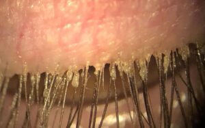
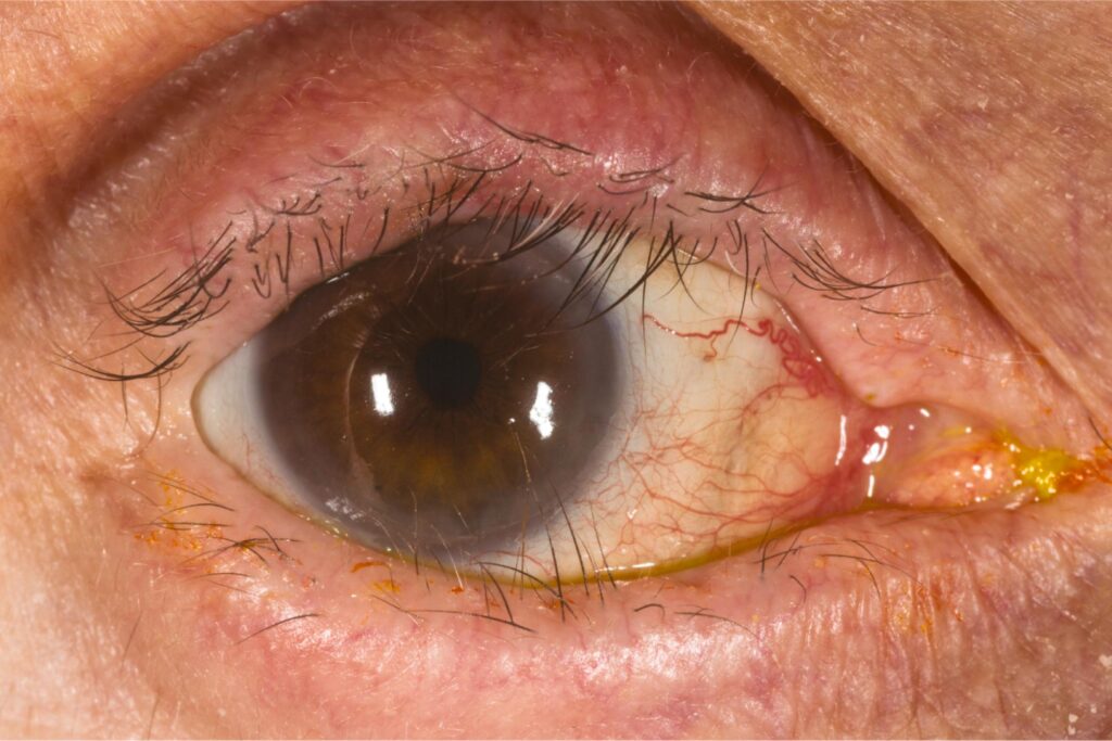
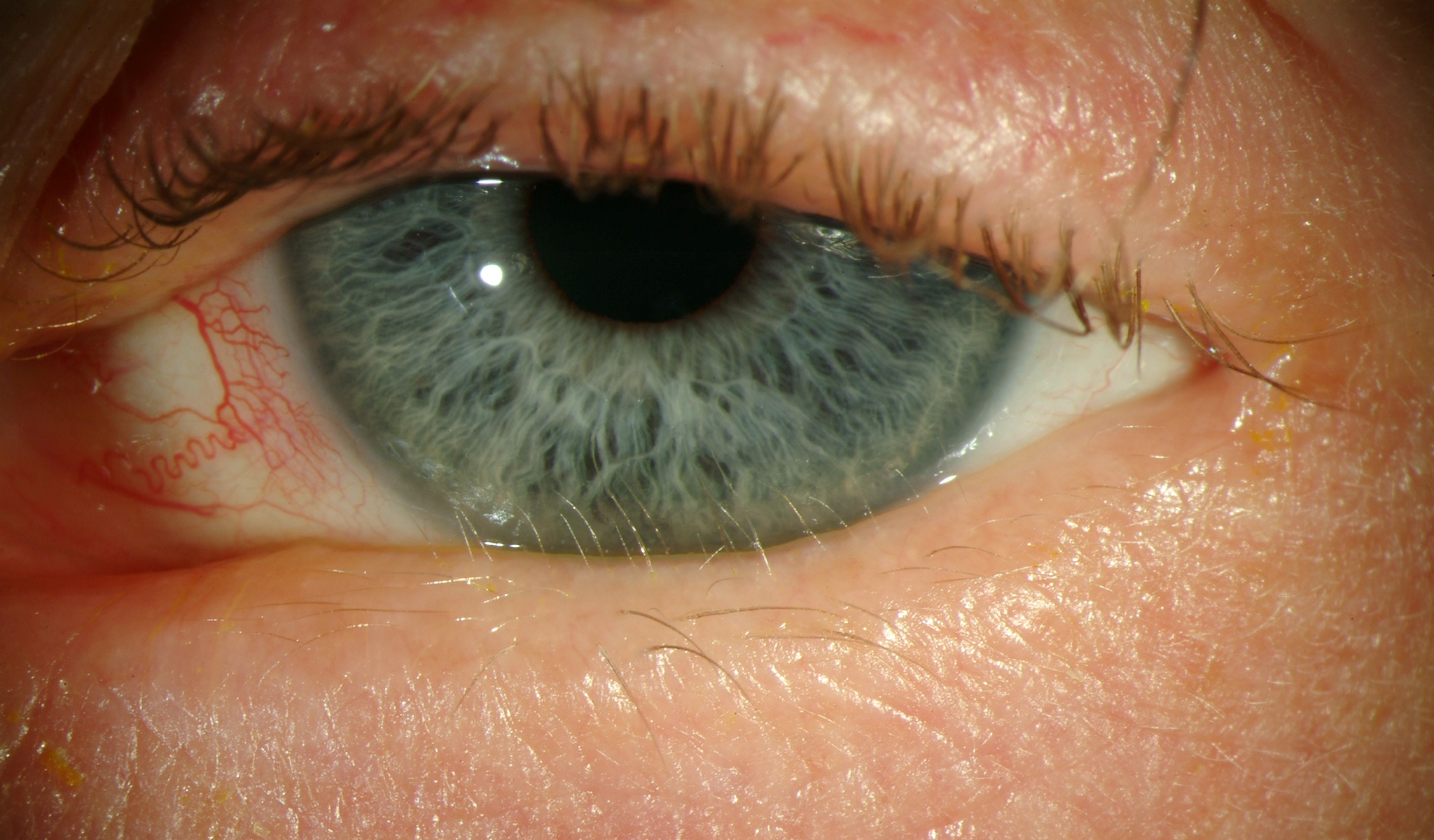
First, locate the gray line, which marks the muscle of Riolan between the lamellae, and next, trace each offending lash back to its exit point on the margin. Lashes posterior to the gray line, at or near meibomian orifices, indicate distichiasis, while lashes anterior to the gray line, within the normal row, indicate trichiasis.
Lash Appearance in Distichiasis vs Trichiasis
Distichiatic lashes usually form a partial or complete second row along the tarsus, and they may stay nearly invisible without high magnification and retroillumination. Trichiatic lashes, by contrast, are mature, pigmented and scattered or segmental.
Corneal Signs and Staining
Fluorescein shows punctate epithelial erosions that follow the lash contact zone, and linear vertical abrasions suggest a lash sweeping across the cornea during blinks. Chronic contact can also cause pannus, stromal scarring and infectious keratitis.
Ruling Out Entropion and Epiblepharon
Inward rotation of the whole lid margin is entropion, which needs a different repair. In epiblepharon, common in East Asian children, a horizontal skin fold rotates lashes against the cornea, so assess lid position before labeling the problem trichiasis.
Cicatricial Disease Behind the Lashes
Posterior margin keratinization, symblepharon or forniceal shortening suggest cicatrizing conjunctivitis, and in that setting both conditions may coexist on the same lid. Mucous membrane pemphigoid deserves active exclusion, because untreated disease progresses and surgery can trigger flares.
Epilation as a Bridge
Forceps epilation gives quick relief in both conditions. However, lashes usually regrow within weeks, and stubby regrowth can irritate more, so a bandage contact lens also protects the cornea while you plan definitive treatment.
Treating Distichiasis vs Trichiasis Differently
Distichiatic follicles lie deep in the tarsus, so the target is the posterior lamella, while trichiatic follicles lie in the anterior lamella, among healthy lashes you want to keep. Therefore, a technique that suits one condition may harm other lid structures.
Follicle Ablation for Trichiasis
For a few lashes, electrolysis or radiofrequency ablation destroys individual follicles, but recurrence is common, so patients may need several sessions. Segmental disease may need wedge excision, while trachomatous trichiasis usually needs bilamellar tarsal rotation.
Lid Splitting for Distichiasis
Many surgeons split the lid at the gray line to separate the lamellae, and then they treat the posterior lamella with cryotherapy or excise the follicles directly. As a result, the normal anterior lashes stay protected, but cryotherapy can damage meibomian glands, depigment skin and worsen cicatricial inflammation.
Counseling in Distichiasis vs Trichiasis
Explain that the misdirected lashes cause the irritation and corneal damage, and tell patients to expect regrowth after epilation and possible repeat procedures. Also, examine relatives of children with congenital distichiasis and ask about leg swelling.
Follow-Up in Distichiasis vs Trichiasis
Review the lid margin with high magnification at each visit, and in addition recheck corneal staining to confirm that the contact zone has healed. Finally, watch cicatricial cases closely, because new aberrant lashes can appear over time.
Pitfalls to Avoid
- Do not treat lashes before tracing their exact origin on the margin
- Never miss entropion or epiblepharon behind apparent trichiasis
- Avoid full-thickness cryotherapy for distichiasis, because it destroys normal lashes
- Do not overlook fine, pale distichiatic lashes at low magnification


Document what you see
Two smartphone imaging tools built for everyday clinic use — one for the slit lamp, one for the fundus.
From Choroida — the team behind this siteReferences
- Bowling B. Kanski’s Clinical Ophthalmology: A Systematic Approach. 8th ed. Edinburgh: Elsevier; 2016.
- Fang J, Dagenais SL, Erickson RP, et al. Mutations in FOXC2 (MFH-1), a forkhead family transcription factor, are responsible for the hereditary lymphedema-distichiasis syndrome. Am J Hum Genet. 2000;67:1382-1388.
- Rajak SN, Collin JR, Burton MJ. Trachomatous trichiasis and its management in endemic countries. Surv Ophthalmol. 2012;57:105-135.
- Vaughn GJ, Dortzbach RK, Sires BS, Lemke BN. Eyelid splitting with excision or microhyfrecation for distichiasis. Arch Ophthalmol. 1997;115:282-284.

Writes and reviews the clinical articles on Educate. Every post is checked against peer-reviewed sources before it goes live.