Blepharitis types fall into two anatomical groups, anterior and posterior.
Anterior disease involves the lash base and the skin of the lid margin, while posterior disease involves the meibomian glands behind the gray line. However, most patients show features of both, so the label describes the dominant pattern.
The Classic Scheme of Blepharitis Types
McCulley and colleagues published a widely used classification of chronic blepharitis in 1982, which separated staphylococcal, seborrheic and meibomian forms, along with mixed patterns. Later, Demodex infestation gained recognition as a distinct anterior cause, so a practical modern scheme lists three anterior types and one posterior type.
Anatomy Behind the Split
The gray line marks the boundary between the two lamellae of the lid margin, with lashes, Zeis glands and Moll glands sitting anterior to it. In contrast, meibomian orifices open posterior to it, just in front of the mucocutaneous junction. As a result, the location of debris and vessels shows which type dominates.
All-fit Slit-Lamp Adapter
Record and share exactly what you see at the slit lamp. One adapter fits any slit lamp or surgical microscope — and any smartphone.
From Choroida — the team behind this siteExamining for Blepharitis Types at the Slit Lamp
First, scan both lid margins with diffuse illumination at low magnification, then ask the patient to look down so you can inspect the upper lash roots. Next, press gently on the central lower lid and watch the secretion (see slit-lamp illumination techniques), and finally instill fluorescein and assess the tear film and inferior cornea.
Staphylococcal Anterior Blepharitis
Hard, brittle fibrinous scales encircle the lash bases, and lifting them may leave small bleeding ulcers on the margin. Chronic cases also show madarosis, trichiasis, poliosis and recurrent hordeola, and in addition the cornea may develop marginal infiltrates or phlyctenules (see staphylococcal blepharitis).
Seborrheic Anterior Blepharitis
Here the scales are soft, greasy and loosely attached to the lashes, and inflammation is usually milder while ulceration is unusual. Because it reflects a skin disorder, look for seborrheic dermatitis on the scalp, brows and nasolabial folds, and it also coexists with meibomian disease in many patients.
Demodex Among Blepharitis Types
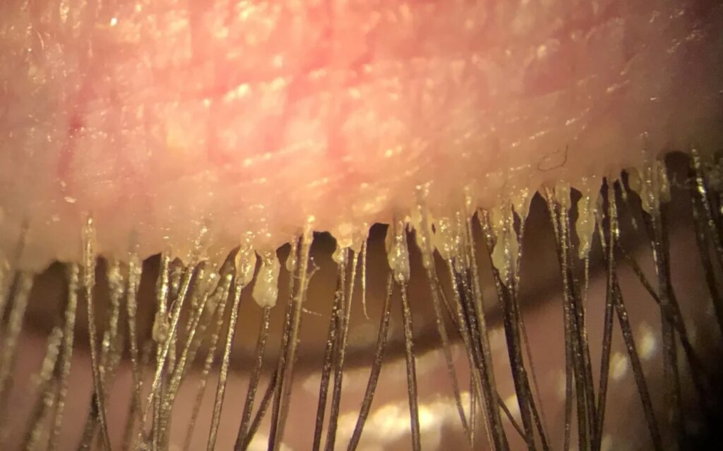
Cylindrical dandruff wraps the lash base like a sleeve, and Gao and colleagues linked this sign to a high prevalence of Demodex in sampled lashes. Patients often report itching along the lid margin, sometimes worse in the morning. For confirmation, epilate a few lashes and view them under a microscope (see Demodex blepharitis).
Posterior Blepharitis and Meibomian Gland Dysfunction
The 2011 international workshop defined meibomian gland dysfunction as a chronic, diffuse gland abnormality, with terminal duct obstruction and altered secretion as its usual features. Signs include capped or pouting orifices, margin telangiectasia and a foamy tear film, while tear breakup time is often short and rosacea frequently coexists.
Grading Meibum in Blepharitis Types
Healthy glands release clear oil with light pressure, but as disease progresses, secretion turns cloudy, then granular, then thick like toothpaste. In addition, some glands express nothing at all, and meibography can then reveal gland dropout that expression alone cannot show.
Symptoms Overlap
Burning, grittiness, morning crusting and fluctuating vision occur in every type, and therefore symptoms rarely classify the disease on their own. Prominent itch points toward Demodex, while evaporative dryness suggests gland disease.
Corneal and Conjunctival Clues
Inferior punctate staining often accompanies meibomian dysfunction. By contrast, staphylococcal hypersensitivity produces peripheral infiltrates with a clear zone from the limbus, and rosacea may also cause corneal vascularization and thinning in severe cases.
Differential Diagnosis
- Sebaceous gland carcinoma, especially unilateral, chronic disease with lash loss
- Allergic contact dermatitis from drops or cosmetics
- Atopic blepharoconjunctivitis
- Herpes simplex blepharitis with vesicles
- Phthiriasis palpebrarum with lice and nits on the lashes
Why Blepharitis Types Guide Treatment
Lid hygiene helps every form, while the rest of the plan follows the dominant type. For example, staphylococcal disease may need topical antibiotic ointment for flares, and lotilaner 0.25% reduced collarettes more than vehicle in the Saturn-2 trial for Demodex.
Meanwhile, gland disease responds to warm compresses, expression and sometimes oral tetracyclines (see stepwise blepharitis treatment). However, the Cochrane review found that most treatments relieve symptoms without a lasting cure.
Mixed Blepharitis Types
Mixed disease is the rule in clinic, and a patient may show cylindrical dandruff and capped glands on the same lid. Then treat the dominant finding first and reassess at follow-up.
Counseling Patients About Blepharitis Types
Explain that blepharitis is chronic and relapsing. Daily lid care controls it, but stopping usually brings symptoms back, and you should also treat the lids before cataract or refractive surgery to reduce surface problems.
Pitfalls to Avoid
- Do not label every red lid margin as simple dry eye
- Never skip a biopsy in persistent unilateral blepharitis
- Do not ignore lash roots when the patient looks straight ahead
- Avoid long courses of steroid drops without monitoring pressure


Document what you see
Two smartphone imaging tools built for everyday clinic use — one for the slit lamp, one for the fundus.
From Choroida — the team behind this siteReferences
- McCulley JP, Dougherty JM, Deneau DG. Classification of chronic blepharitis. Ophthalmology. 1982;89:1173-1180.
- Nelson JD, Shimazaki J, Benitez-del-Castillo JM, et al. The international workshop on meibomian gland dysfunction: report of the definition and classification subcommittee. Invest Ophthalmol Vis Sci. 2011;52:1930-1937.
- Gao YY, Di Pascuale MA, Li W, et al. High prevalence of Demodex in eyelashes with cylindrical dandruff. Invest Ophthalmol Vis Sci. 2005;46:3089-3094.
- Amescua G, Akpek EK, Farid M, et al. Blepharitis Preferred Practice Pattern. Ophthalmology. 2019;126:P56-P93.
- Gaddie IB, Donnenfeld ED, Karpecki P, et al. Lotilaner ophthalmic solution 0.25% for Demodex blepharitis: randomized, vehicle-controlled, multicenter, phase 3 trial (Saturn-2). Ophthalmology. 2023;130:1015-1023.
- Lindsley K, Matsumura S, Hatef E, Akpek EK. Interventions for chronic blepharitis. Cochrane Database Syst Rev. 2012;5:CD005556.

Writes and reviews the clinical articles on Educate. Every post is checked against peer-reviewed sources before it goes live.