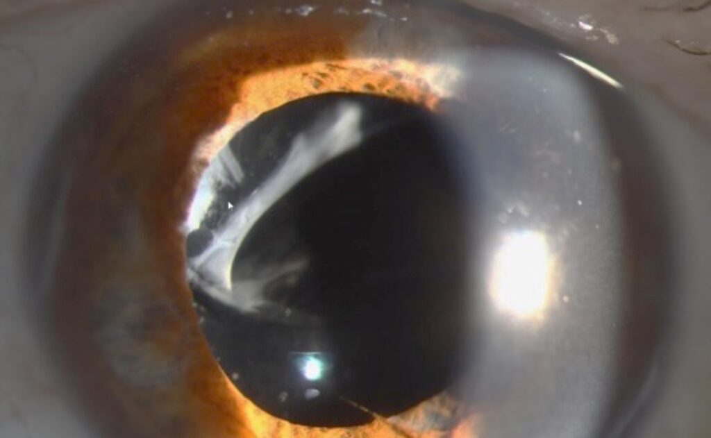
Anterior chamber fibrin is a gelatinous protein coagulum that forms when the blood-aqueous barrier breaks down.
At the slit lamp, it appears as gray translucent strands, a floating clot or a sheet across the pupil.
The sign itself is nonspecific, so the clinical setting decides the diagnosis.
Three settings dominate: HLA-B27 uveitis, the early postoperative eye and endophthalmitis.
Why Anterior Chamber Fibrin Forms
Inflammation opens tight junctions in the iris vessels and ciliary epithelium.
As a result, plasma proteins, including fibrinogen, leak into the aqueous.
Fibrinogen then converts to fibrin, which traps cells and pigment.
What the Examiner Sees
Early fibrin looks like fine strands crossing the pupil or lying on the lens.
Later, it contracts into a dense coagulum or a membrane over the IOL.
In addition, flare is usually intense because protein leakage is heavy.
All-fit Slit-Lamp Adapter
Record and share exactly what you see at the slit lamp. One adapter fits any slit lamp or surgical microscope — and any smartphone.
From Choroida — the team behind this siteAnterior Chamber Fibrin Versus Hypopyon
Fibrin is translucent and gelatinous, while a hypopyon is an opaque white layer of leukocytes.
However, the two often coexist, and fibrin can bind the cells into a sticky mass.
For example, an HLA-B27 hypopyon often looks fibrinous and moves little with head tilt.
By contrast, a Behçet hypopyon tends to shift with gravity in a relatively white eye.
Anterior Chamber Fibrin in HLA-B27 Uveitis
HLA-B27 acute anterior uveitis classically causes sudden, unilateral pain, redness and photophobia.
Fibrin is common in severe attacks, and some eyes develop a fibrinous hypopyon.
Therefore, ask about back pain, bowel symptoms, psoriasis and urethritis (see HLA-B27 workup).
Treating Fibrin in Uveitis
First, start intensive topical corticosteroids, often hourly while awake at the outset.
Next, add a cycloplegic to relieve ciliary spasm and break early synechiae.
In most eyes, the clot then resorbs over days.
However, a dense membrane may need periocular steroid, and the taper should stay slow (see steroid taper).
Complications of Organized Fibrin
Untreated fibrin can organize into a pupillary membrane.
As a result, 360-degree posterior synechiae may cause seclusio pupillae and iris bombé.
Then, angle closure and a sudden pressure rise can follow.
Anterior Chamber Fibrin After Cataract Surgery
Mild fibrin after uneventful surgery is a sterile, expected response in some eyes.
Risk rises in children, diabetics, uveitic eyes and eyes with complicated surgery or iris trauma.
Also, retained lens fragments can drive a persistent fibrinous reaction.
Usually, the patient has mild discomfort and fair vision, and the fundus view stays clear.
Toxic Anterior Segment Syndrome
TASS is a sterile reaction to something introduced into the eye during surgery.
It typically starts within 12 to 24 hours, with diffuse limbus-to-limbus corneal edema.
Fibrin or hypopyon may appear, while pain is often mild and the vitreous stays quiet.
Anterior Chamber Fibrin in Endophthalmitis
Acute postoperative endophthalmitis usually presents a few days to about a week after surgery.
Pain, falling vision and lid edema accompany the fibrin and hypopyon.
Vitreous cells or haze distinguish infection from a purely anterior reaction.
However, dense fibrin may hide the vitreous, so order a B-scan when the view fails.
Red Flags That Suggest Infection
- Pain out of proportion to the surgery
- Vision worse than at the previous visit
- Vitritis, or vitreous opacities on B-scan
- Hypopyon that grows within hours
- Onset after the first postoperative day, with progression on steroids
Managing Suspected Endophthalmitis
Do not wait for a steroid response to test the diagnosis.
Then, perform a vitreous tap and inject intravitreal antibiotics promptly.
In the Endophthalmitis Vitrectomy Study, immediate vitrectomy helped eyes presenting with light perception vision.
Eyes with better vision did similarly well with tap and inject.
Late Fibrin After Surgery
Fibrin or recurrent inflammation weeks to months later should raise doubt.
For example, Cutibacterium acnes can cause a low-grade infection with a white capsular plaque (see chronic postoperative endophthalmitis).
Therefore, consider capsular sampling when inflammation keeps returning after steroid tapers.
Intracameral tPA for Anterior Chamber Fibrin
Intracameral tissue plasminogen activator can dissolve a dense fibrin membrane.
However, its use is off-label, and doses vary between reports.
Also, it can cause hyphema, and fibrin may reform while inflammation persists.
Therefore, combine it with adequate steroid control and warn the patient about bleeding.
Talking With the Patient
Explain that fibrin is a sign of inflammation, and its cause guides treatment.
Patients with uveitis need to know that early treatment of each flare protects the eye.
After surgery, list the warning symptoms (rising pain, falling vision or increasing redness) and ask them to call the same day.
Follow-Up in Anterior Chamber Fibrin
Review severe cases within one to two days.
At each visit, record fibrin extent, cell grade, pressure and synechiae.
Finally, taper steroids only after the fibrin clears and cells settle.
Pitfalls to Avoid
- Do not label a postoperative fibrin reaction sterile without checking the vitreous
- Never stop cycloplegia while fibrin still binds the pupil
- Avoid a fast taper in HLA-B27 attacks with fibrin


Document what you see
Two smartphone imaging tools built for everyday clinic use — one for the slit lamp, one for the fundus.
From Choroida — the team behind this siteReferences
- Endophthalmitis Vitrectomy Study Group. Results of the Endophthalmitis Vitrectomy Study: a randomized trial of immediate vitrectomy and of intravenous antibiotics for the treatment of postoperative bacterial endophthalmitis. Arch Ophthalmol. 1995;113:1479-1496.
- Chang JH, McCluskey PJ, Wakefield D. Acute anterior uveitis and HLA-B27. Surv Ophthalmol. 2005;50:364-388.
- Mamalis N, Edelhauser HF, Dawson DG, et al. Toxic anterior segment syndrome. J Cataract Refract Surg. 2006;32:324-333.
- Jabs DA, Nussenblatt RB, Rosenbaum JT; SUN Working Group. Standardization of uveitis nomenclature for reporting clinical data. Am J Ophthalmol. 2005;140:509-516.

Writes and reviews the clinical articles on Educate. Every post is checked against peer-reviewed sources before it goes live.