Hudson-Stahli line is a horizontal line of iron in the basal corneal epithelium, usually at the junction of the lower and middle thirds of the cornea.
It is common in older adults and it is harmless, so recognizing it prevents unnecessary work-up and avoids confusion with pathological lines.
What the Hudson-Stahli Line Looks Like
The line is fine, yellow-brown, and horizontal, and it often runs along the level of lid closure. It may be complete across the cornea or only partial, and both eyes usually show the same pattern. Because it lies in the epithelium, it does not stain with fluorescein.
Why the Hudson-Stahli Line Forms
Iron deposits in the basal epithelial cells at the line, and tear film pooling and the mechanics of the blink are thought to concentrate iron there. Also, the line becomes more frequent with age, which fits a slow, cumulative process.
All-fit Slit-Lamp Adapter
Record and share exactly what you see at the slit lamp. One adapter fits any slit lamp or surgical microscope — and any smartphone.
From Choroida — the team behind this siteHudson-Stahli Line Versus Other Iron Lines
Position and setting separate the iron lines, as the Fleischer ring surrounds a keratoconic cone and Stocker’s line sits at the head of a pterygium. Ferry’s line lies in front of a filtering bleb, and a Kayser-Fleischer ring lies in Descemet membrane at the limbus (see Kayser-Fleischer ring). Therefore, a horizontal line in an older patient with a normal cornea is almost always the Hudson-Stahli line.
What to Do When You See a Hudson-Stahli Line
No treatment is needed, so reassure the patient and record the finding so that a later examiner does not mistake it for a new lesion. If the pattern looks unusual, look for a cone, a bleb or a foreign body (see corneal iron lines).
Prevalence and Age
The line is uncommon in children and becomes frequent in later life, and it usually appears in both eyes in patients who have it. Therefore, a one-sided line should prompt a closer look for another cause.
Appearance Under Different Lighting
Under white light, the line looks like a faint brown streak, while under cobalt blue light it appears dark and easier to follow. Also, a broad beam at an angle shows its full length against the pale cornea.
Symptoms
The line causes no symptoms and no visual loss, so if the patient complains of discomfort, look for another cause, such as dry eye or blepharitis (see dry eye disease).
Pitfalls With Staining
A line that stains with fluorescein points to abrasion or a linear erosion and not to an iron line, and an edge of a contact lens can leave a mark that mimics the pattern. Because iron lines sit within the epithelium, they do not take up dye.
Distinguishing It From a Kayser-Fleischer Ring
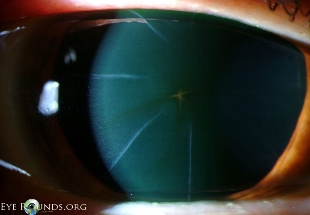
The Kayser-Fleischer ring is greenish-brown, lies in Descemet membrane, and forms a circle at the limbus. The Hudson-Stahli line is horizontal and lies in the epithelium, so test for copper only when you see a ring at the limbus.
Documentation
Note the line in the chart as bilateral and benign, so that future examiners will not mistake it for a new lesion.
Clinical Setting
Most patients come with an unrelated complaint, and the line is an incidental finding, and patients often have the line in both eyes without any other corneal disease.
Why Recognition Helps
Recognizing the line avoids unnecessary tests and spares the patient worry, and because other iron lines point to disease, learning the pattern helps you name them correctly. Also, when you can name the line at once, you save time and avoid needless tests.
Teaching Points for Trainees
Ask trainees to find the line with cobalt blue light in older patients. Then ask them to name three other iron lines and the diseases they accompany, and remind them that location is the key to the diagnosis.
Quick Comparison Table in Words
The Hudson-Stahli line is horizontal and inferior, Stocker’s line is at the pterygium head, and the Fleischer ring surrounds the cone. Ferry’s line sits in front of a bleb, and the Coats ring marks a past iron foreign body, so each line is a map of a local corneal change.
Hudson-Stahli Line and Other Linear Corneal Findings
Several linear findings can look like an iron line, and map-dot-fingerprint dystrophy produces curved lines and dots in the epithelium (see map-dot-fingerprint dystrophy). Scratches from a foreign body stain with fluorescein and cause pain, so the history and the stain test usually settle the question.
Hudson-Stahli Line and Tear Film Dynamics
The line lies near the level where the lids meet the cornea during the blink. Tear film dynamics at this level may explain why iron settles there, and the same position in both eyes supports a mechanical cause.


Document what you see
Two smartphone imaging tools built for everyday clinic use — one for the slit lamp, one for the fundus.
From Choroida — the team behind this siteReferences
- Gass JDM. The iron lines of the superficial cornea: Hudson-Stahli line, Stocker’s line, and Fleischer’s ring. Arch Ophthalmol. 1964;71:348-358.
- Barraquer-Somers E, Chan CC, Green WR. Corneal epithelial iron deposition. Ophthalmology. 1983;90:1012-1017.
- Mannis MJ, Holland EJ, eds. Cornea. 5th ed. Elsevier; 2022.
- Kanski JJ, Bowling B. Clinical Ophthalmology: A Systematic Approach. 8th ed. Elsevier; 2016.
Test yourself
A few questions straight from this article.
-
Where is the Hudson-Stahli line typically located in the cornea?
The article defines the Hudson-Stahli line as a horizontal line of iron in the basal corneal epithelium, usually at the junction of the lower and middle thirds of the cornea. -
Which description fits the typical appearance of a Hudson-Stahli line?
The article describes the line as fine, yellow-brown and horizontal, often running along the level of lid closure. -
How does a Hudson-Stahli line behave with fluorescein, and why?
Because the line lies in the epithelium, it does not stain with fluorescein. A line that stains suggests abrasion or linear erosion instead. -
What is thought to concentrate iron in the Hudson-Stahli line?
The article states that iron deposits in basal epithelial cells and that tear film pooling and blink mechanics are thought to concentrate iron there. -
Which iron line surrounds the cone in keratoconus, unlike the Hudson-Stahli line?
The article notes that the Fleischer ring surrounds a keratoconic cone, while the Hudson-Stahli line is horizontal and inferior in a normal cornea. -
Which iron line sits at the head of a pterygium?
Position and setting separate the iron lines. Stocker's line sits at the head of a pterygium, whereas Ferry's line lies in front of a filtering bleb. -
When should copper testing be considered in a patient with a Hudson-Stahli line-like finding?
The Kayser-Fleischer ring is greenish-brown, lies in Descemet membrane and forms a circle at the limbus. Copper testing is only needed when such a ring is seen. -
What is the appropriate management of a Hudson-Stahli line?
The line is harmless, so no treatment is needed. Reassure the patient and record it so a later examiner does not mistake it for a new lesion. -
A patient has a one-sided Hudson-Stahli-like line. What does the article advise?
The line usually appears in both eyes, so a one-sided line should prompt a closer look for another cause. -
Which light makes a Hudson-Stahli line dark and easier to follow?
Under white light the line is a faint brown streak, while under cobalt blue light it appears dark and easier to follow.

Writes and reviews the clinical articles on Educate. Every post is checked against peer-reviewed sources before it goes live.