Capsulorhexis is a continuous circular tear of the anterior lens capsule, made at the start of phacoemulsification.
A well-centered, round opening of the right size supports the intraocular lens and protects the capsular bag. Therefore, it is one of the most important steps of the operation.
What Capsulorhexis Achieves
Neuhann and Gimbel introduced the continuous curvilinear technique in the 1980s to replace the can-opener capsulotomy. The continuous edge resists radial tears, so the bag stays strong through hydrodissection and nuclear rotation.
It also allows the lens to sit in the bag, with the optic covered by the capsule edge. Because the bag holds the implant for life, a clean opening protects the long-term result.
Preparing for Capsulorhexis
Dilate the pupil well and fill the anterior chamber with a cohesive viscoelastic. The viscoelastic flattens the anterior capsule and holds back the posterior pressure.
Also, check that the incision is the right size and shape, since leakage destabilizes the chamber. Then choose a cystotome or capsulorhexis forceps that you can control.
All-fit Slit-Lamp Adapter
Record and share exactly what you see at the slit lamp. One adapter fits any slit lamp or surgical microscope — and any smartphone.
From Choroida — the team behind this siteTechnique of Capsulorhexis
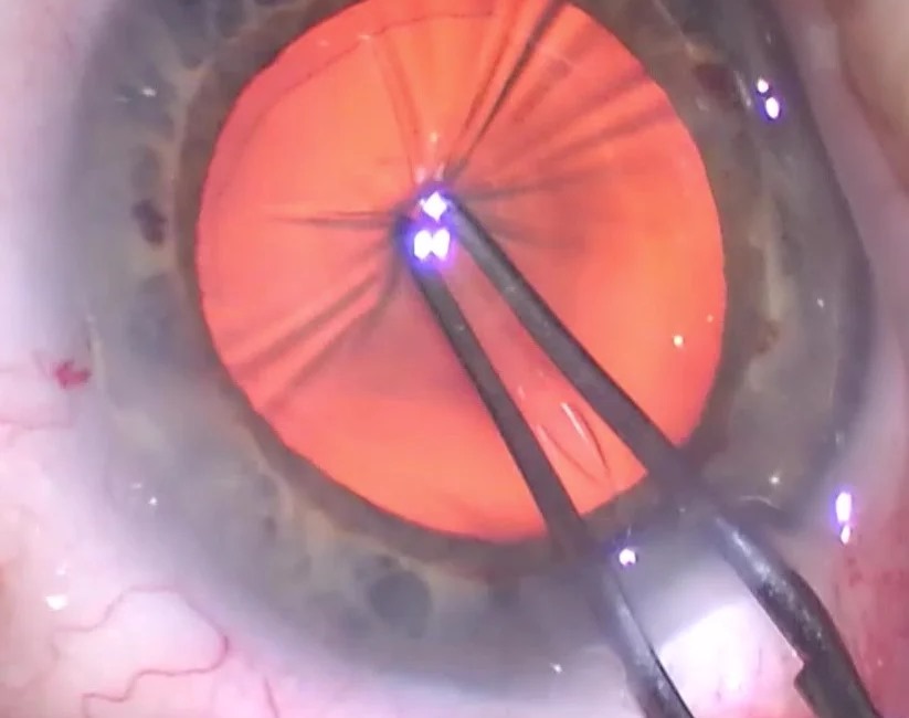
Puncture the capsule centrally and lift a small flap, then fold the flap over and drag the leading edge in a circular path around the pupil. Regrasp the flap close to the tear edge as you go, which gives you better control. Finally, complete the circle by overlapping the start of the tear and take the flap out of the way.
Ideal Size and Position
Aim for an opening of about 5 to 5.5 millimeters, centered on the visual axis. The edge should overlap the optic of the lens by a small margin all around.
A small opening raises the risk of capsular contraction and makes nuclear delivery harder (see anterior capsular contraction). In contrast, a large opening allows the lens to decenter and leaves the optic partly uncovered.
Capsulorhexis in White Cataract
A white cataract has no red reflex, so the capsule is hard to see, but trypan blue stains the anterior capsule and makes the edge visible. In an intumescent lens, the pressure pushes the tear outward, so puncture and aspirate the liquefied cortex first (see white cataracts), and use a small starting opening and enlarge it slowly.
Rescuing a Radial Tear
A tear that runs toward the periphery needs a pause. Fill the chamber with cohesive viscoelastic to flatten the capsule, and then redirect the tear toward the center.
If the tear continues, make a new flap and tear back from the opposite side. Because a tear that reaches the equator can extend to the posterior capsule, convert to a low-stress technique for the rest of the case.
Capsulorhexis With Weak Zonules and Small Pupils
Pseudoexfoliation and trauma weaken the zonules, so use gentle, short movements. Capsule hooks and tension rings support the bag during surgery (see capsular tension ring).
In a small pupil, use iris hooks or a ring to expand the pupil before you start. Also, plan a larger incision and a staged approach when the view is poor.
After Surgery
The capsule edge usually stays visible, and a ring of fibrosis may form over time, while in some eyes the opening shrinks, which is called capsular phimosis. Posterior capsule opacification may need a laser opening later (see YAG laser capsulotomy). Also, check the edge position at follow-up, since a decentered opening can explain optical symptoms.
Instruments and Incisions
Capsulorhexis forceps and a cystotome both work, and each has strengths. Forceps give better control in a flat chamber, because they grasp the edge directly. A cystotome needs less space and is easier through a small paracentesis.
Also, a well-sized main incision lets you move the instrument freely without leakage. Therefore, choose the tool that fits the case and your habits.
Learning Curve and Training
Capsulorhexis is a skill that improves with deliberate practice, and wet labs and simulators allow trainees to rehearse the flap, the regrasp and the circle. Also, reviewing video of your own cases shows errors in size and centration. Because small tears teach the most, ask a mentor to review a few cases with you.
Pediatric and Special Cases
The pediatric capsule is thin and elastic, so the tear tends to run outward. Use a viscoelastic with high retention and a smaller initial tear, and consider posterior capsulorhexis and vitrectomy in young children to prevent opacity. In adults with posterior polar cataract, keep a smaller opening and avoid hydrodissection.
Documentation
Record the size, the centration and any tear in the operative note, and note whether the edge covers the optic fully. Because the edge position affects later laser capsulotomy, a clear note helps.


Document what you see
Two smartphone imaging tools built for everyday clinic use — one for the slit lamp, one for the fundus.
From Choroida — the team behind this siteReferences
- Gimbel HV, Neuhann T. Development, advantages, and methods of the continuous circular capsulorhexis technique. J Cataract Refract Surg. 1990;16:31-37.
- Assia EI, Apple DJ, Barden A, et al. An experimental study comparing various anterior capsulectomy techniques. Arch Ophthalmol. 1991;109:642-647.
- Little BC, Smith JH, Packer M. Little capsulorhexis tear-out rescue. J Cataract Refract Surg. 2006;32:1420-1422.
- Marques FF, Marques DM, Osher RH, Osher JM. Fate of anterior capsule tears during cataract surgery. J Cataract Refract Surg. 2006;32:1638-1642.
Test yourself
A few questions straight from this article.
-
In cataract surgery, capsulorhexis is a continuous circular tear of which structure?
Capsulorhexis is a continuous circular tear of the anterior lens capsule made at the start of phacoemulsification. -
Why does a continuous curvilinear capsulorhexis keep the capsular bag stronger than a can-opener capsulotomy?
The continuous edge resists radial tears, so the bag stays strong through hydrodissection and nuclear rotation. -
Before starting a capsulorhexis, what does filling the anterior chamber with cohesive viscoelastic achieve?
Cohesive viscoelastic flattens the anterior capsule and holds back posterior pressure, which keeps the tear under control. -
What capsulorhexis size and position is recommended in routine cataract surgery?
An opening of about 5 to 5.5 mm centred on the visual axis lets the edge overlap the lens optic by a small margin all around. -
Which problem is associated with a capsulorhexis that is too small?
A small opening raises the risk of capsular contraction and makes nuclear delivery harder, whereas a large one lets the lens decenter. -
In a white cataract with no red reflex, how is the anterior capsule made visible for capsulorhexis?
Without a red reflex the capsule is hard to see, and trypan blue stains the anterior capsule so the tear edge becomes visible. -
In an intumescent white cataract, what step reduces the outward pull on the capsulorhexis tear?
Intralenticular pressure pushes the tear outward, so the liquefied cortex is aspirated first and a small opening is enlarged slowly. -
During capsulorhexis, a tear starts running toward the periphery. What is the first rescue step?
Pause, flatten the capsule with cohesive viscoelastic and redirect the tear toward the centre; if it continues, tear back from the opposite side. -
In pseudoexfoliation with weak zonules, what supports the capsular bag during capsulorhexis and surgery?
Pseudoexfoliation and trauma weaken the zonules, so gentle short movements are used and capsule hooks and tension rings support the bag. -
After cataract surgery, the capsulorhexis opening shrinks in some eyes. What is this called?
The capsule edge usually stays visible and may fibrose; when the opening shrinks over time, this is called capsular phimosis.

Writes and reviews the clinical articles on Educate. Every post is checked against peer-reviewed sources before it goes live.