Retinitis vs choroiditis is the first layer question to answer when a white or yellow fundus lesion appears with uveitis.
Retinitis starts in the neurosensory retina, while choroiditis starts in the vascular layer beneath the RPE. Because each layer attracts different organisms, locating the lesion shortens the infectious workup.
Why Layer Location Guides Retinitis vs Choroiditis
The SUN Working Group classifies posterior uveitis by the primary tissue involved, and retina and choroid differ in blood supply, immune barriers and typical pathogens. Therefore, naming the layer turns a vague white spot into a focused differential.
Lesion Color and Border in Retinitis vs Choroiditis
Active retinitis looks white, opaque and fluffy, with edges that fade into normal retina. Choroidal lesions, in contrast, look yellow or cream and appear deeper, with usually sharper borders, while the overlying retina often stays transparent.
Fundus Explorer Pro
Photograph the retinal findings described here with the phone already in your pocket — 22 D optics and built-in illumination in one handheld unit.
From Choroida — the team behind this siteRetinal Vessels in Retinitis vs Choroiditis
Next, follow the retinal vessels across the lesion. In retinitis, swollen inner retina hides the vessels running through the white area, whereas in choroiditis, retinal vessels pass over the lesion clearly because the inflammation lies beneath them. Sheathing, occlusion and intraretinal hemorrhage also favor retinitis, especially in herpetic and CMV disease.
Vitreous Reaction
Retinitis sheds cells directly into the vitreous, so dense vitritis is typical. For example, toxoplasmosis can produce the classic “headlight in the fog” appearance.
Choroiditis often causes milder vitritis because the RPE separates the focus from the gel. However, immunosuppression blunts vitritis in both groups, so a quiet vitreous never excludes retinitis.
OCT in Retinitis vs Choroiditis
OCT confirms the layer when the fundus looks ambiguous. Active retinitis shows thickened, hyperreflective retina with lost lamination and posterior shadowing. In choroiditis, the retina may look intact while EDI-OCT shows hyporeflective choroidal infiltrates and choroidal thickening (see choroidal thickness on EDI-OCT).
Fluorescein Angiography Findings
Fluorescein mainly reports retinal vessel and RPE behavior. Active retinitis typically blocks early and stains or leaks at its margins late, often with adjacent vasculitis, but deep choroidal lesions can follow a similar pattern, so FA alone rarely settles the layer.
ICG Angiography in Choroiditis
Indocyanine green penetrates the RPE and images the choroidal stroma well. Choroidal granulomas appear as hypofluorescent dark dots that persist into the late phase, and ICG often reveals more lesions than ophthalmoscopy, for example in sarcoidosis, tuberculosis and birdshot chorioretinopathy.
Infectious Retinitis Differential
Once the lesion sits in the retina, a short infectious list follows. Toxoplasmosis often lies beside an old pigmented scar, while CMV retinitis occurs mainly in immunocompromised patients.
Acute retinal necrosis from HSV or VZV spreads peripherally and fast, and syphilis can cause punctate inner retinitis. Also consider progressive outer retinal necrosis when profound immunosuppression meets minimal inflammation.
Infectious Choroiditis Differential
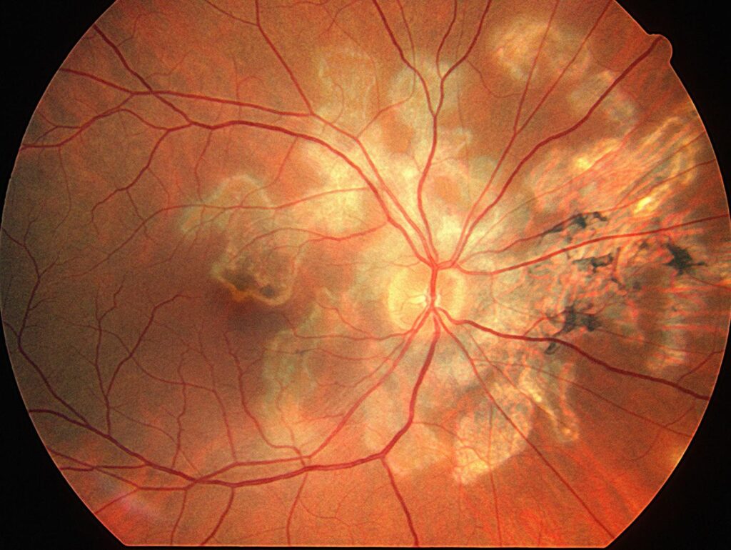
Choroidal lesions suggest a different set of organisms. Tuberculosis causes choroidal tubercles, granulomas and serpiginous-like choroiditis.
Syphilis may produce acute syphilitic posterior placoid chorioretinitis, as Gass and colleagues described. In addition, Pneumocystis, Cryptococcus and endogenous fungi can seed the choroid in immunocompromised hosts.
Noninfectious Mimics in Retinitis vs Choroiditis
Most noninfectious disease favors the choroid, including sarcoidosis, birdshot chorioretinopathy, VKH and multifocal choroiditis. Behçet disease is the main exception, because occlusive retinal vasculitis produces patches of retinitis. Finally, intraocular lymphoma forms sub-RPE infiltrates that mimic choroiditis in older adults.
When Lesions Cross Layers
Toxoplasmosis begins in the retina and then involves the adjacent choroid, hence the term retinochoroiditis. Conversely, Candida starts in the choroid and then breaks through the retina into the vitreous, so document where the lesion began and how it evolved on serial photographs.
Testing After Retinitis vs Choroiditis Localization
For necrotizing retinitis, aqueous or vitreous PCR for HSV, VZV, CMV and Toxoplasma gives the fastest answer. For choroiditis, an interferon-gamma release assay, chest imaging and syphilis serology are reasonable first steps, and syphilis testing suits both groups because the disease mimics almost every pattern.
Treatment Timing and Counseling
Herpetic retinitis can destroy retina within days. As a result, start antiviral therapy when clinical suspicion is strong, without waiting for PCR results.
Choroiditis usually allows more time, although corticosteroids should still wait until infection is reasonably excluded. Then explain why tests come before steroids, and ask patients to report new floaters or vision loss promptly.
Follow-Up in Retinitis vs Choroiditis
Serial widefield photos show whether borders advance or consolidate. Healing retinitis turns granular and then atrophic, while choroidal lesions fade, pigment or leave punched-out scars, and repeat OCT can detect macular edema, choroidal neovascularization and retinal detachment.
Pitfalls to Avoid
- Do not label a deep yellow lesion retinitis because vitritis is dense
- Never give steroids alone before excluding infection
- Do not trust a quiet vitreous in immunosuppressed patients
- Always test for syphilis in posterior uveitis


Document what you see
Two smartphone imaging tools built for everyday clinic use — one for the slit lamp, one for the fundus.
From Choroida — the team behind this siteReferences
- Jabs DA, Nussenblatt RB, Rosenbaum JT; Standardization of Uveitis Nomenclature (SUN) Working Group. Standardization of uveitis nomenclature for reporting clinical data. Results of the First International Workshop. Am J Ophthalmol. 2005;140:509-516.
- Holland GN. Ocular toxoplasmosis: a global reassessment. Part II: disease manifestations and management. Am J Ophthalmol. 2004;137:1-17.
- Gass JD, Braunstein RA, Chenoweth RG. Acute syphilitic posterior placoid chorioretinitis. Ophthalmology. 1990;97:1288-1297.
- Herbort CP. Fluorescein and indocyanine green angiography for uveitis. Middle East Afr J Ophthalmol. 2009;16:168-187.
- American Academy of Ophthalmology. Basic and Clinical Science Course, Section 9: Uveitis and Ocular Inflammation. San Francisco: AAO; 2023.

Writes and reviews the clinical articles on Educate. Every post is checked against peer-reviewed sources before it goes live.