Hypertensive optic neuropathy is optic disc swelling caused by severe, usually acute, elevation of blood pressure.
Most patients have bilateral findings, although the two discs may look asymmetric. Therefore, a swollen disc with a very high reading needs same-day medical assessment.
What Hypertensive Optic Neuropathy Means
The term describes disc edema within the spectrum of malignant hypertension, and it often accompanies hypertensive retinopathy and choroidopathy. However, the disc can swell with few retinal signs, and classic Keith-Wagener-Barker grading placed disc swelling in group 4, which carried the worst survival.
Why the Disc Swells
The mechanism remains debated, and Hayreh and colleagues attributed the swelling mainly to optic nerve head ischemia in experimental malignant hypertension. In addition, raised intracranial pressure may contribute in some patients, and several processes may therefore overlap in one eye.
Fundus Explorer Pro
Photograph the retinal findings described here with the phone already in your pocket — 22 D optics and built-in illumination in one handheld unit.
From Choroida — the team behind this siteSymptoms That Raise Concern
Many patients report blurred vision, headache or both, while others notice little because central acuity can stay good until the macula becomes involved. Meanwhile, confusion, seizures, chest pain or dyspnea suggest wider organ damage. Any of these complaints turns the visit into a medical emergency.
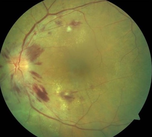
Fundus Signs of Hypertensive Optic Neuropathy
Dilated examination usually shows more than a swollen disc, so look for these associated findings:
- Hyperemic disc edema with blurred margins
- Flame hemorrhages and cotton wool spots
- Macular star from lipid exudation
- Serous retinal detachment
- Elschnig spots or Siegrist streaks, which indicate choroidal ischemia
- Arteriolar narrowing and arteriovenous nicking, which suggest chronic disease
Measuring Blood Pressure
Measure blood pressure in every patient with bilateral disc swelling, use a correctly sized cuff, and then repeat the reading if it looks high. Guidelines commonly use 180/120 mmHg as the threshold for severe hypertension, although a lower reading can still cause damage in pregnancy.
Hypertensive Optic Neuropathy as an Emergency
Acute disc swelling with severe hypertension signals possible target organ damage, and the brain, heart and kidneys may all be at risk. Therefore, arrange direct transfer to emergency care, send your findings with the patient, and do not book a routine outpatient review.
Separating Hypertensive Optic Neuropathy From Papilledema
Papilledema means disc swelling from raised intracranial pressure, and it can look identical (see is every swollen disc papilledema?). Therefore, blood pressure alone should not close the diagnosis, and neuroimaging helps exclude a mass, hemorrhage or venous sinus thrombosis. Lumbar puncture may follow normal imaging when the cause remains unclear.
Other Causes to Exclude
A broader framework helps when the picture does not fit (see optic disc swelling causes), so consider these alternatives:
- NAION, which is usually unilateral with an altitudinal defect
- Arteritic ischemia, especially with jaw claudication
- Optic neuritis, which typically causes painful eye movement
- Neuroretinitis, which also produces a macular star
- Central retinal vein occlusion and disc drusen
Clues From Laterality
Bilateral swelling favors a systemic or intracranial cause, whereas unilateral swelling with a relative afferent pupillary defect points toward a primary optic nerve process.
Imaging the Eye
OCT documents nerve fiber layer thickening, subretinal fluid and macular involvement, and it also provides a baseline for later ganglion cell thinning. Fluorescein angiography may show disc leakage and delayed choroidal filling, but angiography should never delay blood pressure control.
Systemic Workup in Hypertensive Optic Neuropathy
The medical team usually checks renal function, urinalysis, ECG and cardiac markers. Younger patients deserve a search for secondary causes, including renal disease and pheochromocytoma, and in pregnancy, preeclampsia must be considered first.
Lowering Pressure Safely
Physicians usually use titratable intravenous agents, and the ACC/AHA guideline generally advises reducing pressure by no more than 25% within the first hour. Then, targets are approached stepwise over the next 24 to 48 hours.
Chronic hypertension shifts autoregulation, so abrupt lowering can cause optic nerve ischemia. Therefore, share your disc findings with the treating physician, and then recheck vision during treatment.
What Treatment Cannot Do
Blood pressure control lets swelling and exudates resolve, often over weeks to months, but it cannot restore axons that have already died. Optic atrophy and permanent field loss can therefore persist (see pale disc after injury).
Counseling and Follow-Up in Hypertensive Optic Neuropathy
Explain that the eye findings reflect a body-wide emergency. Many patients regain useful vision, although recovery varies. Reassess acuity, fields, disc appearance and OCT after discharge, and also stress long-term medication adherence, because recurrent surges threaten the eyes, brain, heart and kidneys.
Pitfalls to Avoid
- Do not call bilateral disc swelling papilledema before checking blood pressure
- Avoid attributing swelling to hypertension when focal neurological signs remain unexplained
- Never overlook pregnancy in a young woman with disc swelling
- Do not advise rapid pressure lowering at home
- Avoid assuming normal acuity means low risk


Document what you see
Two smartphone imaging tools built for everyday clinic use — one for the slit lamp, one for the fundus.
From Choroida — the team behind this siteReferences
- Hayreh SS, Servais GE, Virdi PS. Fundus lesions in malignant hypertension. V. Hypertensive optic neuropathy. Ophthalmology. 1986;93:74-87.
- Wong TY, Mitchell P. Hypertensive retinopathy. N Engl J Med. 2004;351:2310-2317.
- Keith NM, Wagener HP, Barker NW. Some different types of essential hypertension: their course and prognosis. Am J Med Sci. 1939;197:332-343.
- Whelton PK, Carey RM, Aronow WS, et al. 2017 ACC/AHA guideline for the prevention, detection, evaluation, and management of high blood pressure in adults. Hypertension. 2018;71:e13-e115.
- Williams B, Mancia G, Spiering W, et al. 2018 ESC/ESH guidelines for the management of arterial hypertension. Eur Heart J. 2018;39:3021-3104.
Test yourself
A few questions straight from this article.
-
What is hypertensive optic neuropathy?
The article defines hypertensive optic neuropathy as optic disc swelling caused by severe, usually acute, elevation of blood pressure. It sits within the spectrum of malignant hypertension. -
Which mechanism did Hayreh and colleagues mainly propose for disc swelling in experimental malignant hypertension?
The article states the mechanism is debated, but Hayreh and colleagues attributed the swelling mainly to optic nerve head ischemia. Raised intracranial pressure may also contribute in some patients. -
In hypertensive optic neuropathy, why may a patient report few visual symptoms despite a swollen disc?
The article notes that many patients notice little because central acuity can stay good until the macula is involved. It also warns against assuming normal acuity means low risk. -
Which finding in hypertensive optic neuropathy indicates choroidal ischemia?
Elschnig spots and Siegrist streaks indicate choroidal ischemia. Arteriolar narrowing and arteriovenous nicking instead suggest chronic disease. -
What blood pressure threshold do guidelines commonly use for severe hypertension in a patient with hypertensive optic neuropathy?
The article says guidelines commonly use 180/120 mmHg as the threshold for severe hypertension. A lower reading can still cause damage in pregnancy. -
A patient has acute disc swelling from hypertensive optic neuropathy with severe hypertension. What is the appropriate disposition?
Acute disc swelling with severe hypertension signals possible target organ damage to the brain, heart and kidneys. The article advises direct transfer to emergency care and not a routine outpatient review. -
Why should blood pressure alone not close the diagnosis in a patient with hypertensive optic neuropathy and a swollen disc?
Papilledema from raised intracranial pressure can look identical to hypertensive disc swelling. Neuroimaging helps exclude a mass, hemorrhage or venous sinus thrombosis, and lumbar puncture may follow if the cause remains unclear. -
What laterality clue favors a primary optic nerve process over a systemic cause of disc swelling?
Bilateral swelling favors a systemic or intracranial cause. Unilateral swelling with a relative afferent pupillary defect points toward a primary optic nerve process. -
How quickly does the ACC/AHA guideline generally advise lowering blood pressure in hypertensive optic neuropathy during the first hour?
The article says the ACC/AHA guideline generally advises reducing pressure by no more than 25% within the first hour. Targets are then approached stepwise over 24 to 48 hours. -
Why can abrupt blood pressure lowering harm a patient with hypertensive optic neuropathy?
Chronic hypertension shifts autoregulation, so abrupt lowering can cause optic nerve ischemia. The article advises sharing disc findings with the treating physician and rechecking vision during treatment.

Writes and reviews the clinical articles on Educate. Every post is checked against peer-reviewed sources before it goes live.