VKH stages describe the predictable course of Vogt-Koyanagi-Harada disease, a bilateral granulomatous panuveitis that targets melanocytes.
Moorthy and colleagues divided this course into prodromal, acute uveitic, convalescent and chronic recurrent phases.
Recognizing the phase helps you choose tests, time treatment and anticipate complications.
For a broader overview, see our review of Vogt-Koyanagi-Harada disease.
How VKH Stages Are Defined
The phases reflect an immune attack on melanocytes in the choroid, skin, meninges and inner ear.
Therefore, early signs are inflammatory, while later signs are depigmenting.
The phases often overlap, and prompt treatment can change their course.
Prodromal Phase Symptoms
First, the prodrome usually lasts a few days and resembles a viral illness.
Patients report headache, fever, nausea, orbital pain and neck stiffness.
Some also notice tinnitus, vertigo or reduced hearing.
Scalp and hair sensitivity to touch is a typical, often missed complaint.
Fundus Explorer Pro
Photograph the retinal findings described here with the phone already in your pocket — 22 D optics and built-in illumination in one handheld unit.
From Choroida — the team behind this siteProdromal Findings in VKH Stages
The eye examination may still look normal at this point.
However, lumbar puncture can show a lymphocytic pleocytosis in the cerebrospinal fluid.
Because patients rarely link headache to blurred vision, ask about these symptoms directly.
Acute Uveitic Phase on Fundus Exam
Blurred vision in both eyes follows within days, although the second eye may lag briefly.
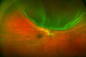
The fundus shows optic disc hyperemia, choroidal thickening and multiple serous retinal detachments.
In severe cases, these detachments coalesce into a bullous exudative detachment.
OCT in the Acute Uveitic Phase
OCT shows subretinal fluid divided by septa or membranous strands.
Undulating RPE folds and a markedly thickened choroid on enhanced depth imaging are also typical.
As a result, OCT confirms the clinical picture quickly and sets a baseline for monitoring.
Angiography in Early VKH Stages
Fluorescein angiography shows multiple pinpoint leaks at the level of the RPE.
Then, dye pools within the serous detachments on late frames.
Indocyanine green angiography adds hypofluorescent dark dots and fuzzy choroidal vessels.
Because these dark dots often persist after the fluid clears, they help detect subclinical choroiditis.
Anterior Segment in Acute Disease
The anterior chamber may show only mild cells early.
However, ciliary body swelling can shallow the chamber and cause a sudden myopic shift.
This picture can mimic primary acute angle closure.
Therefore, treat the inflammation, because miotics may worsen this type of closure.
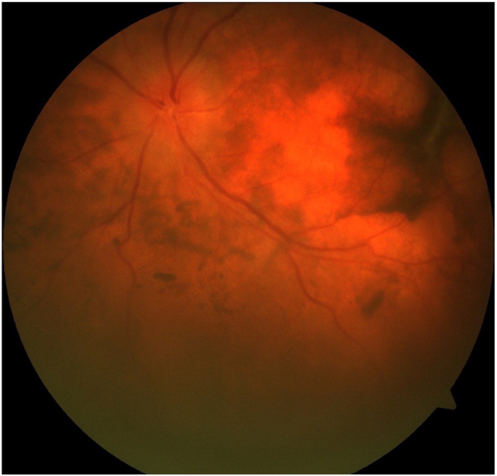
Convalescent Phase Depigmentation
Weeks to months later, inflammation settles and depigmentation appears.
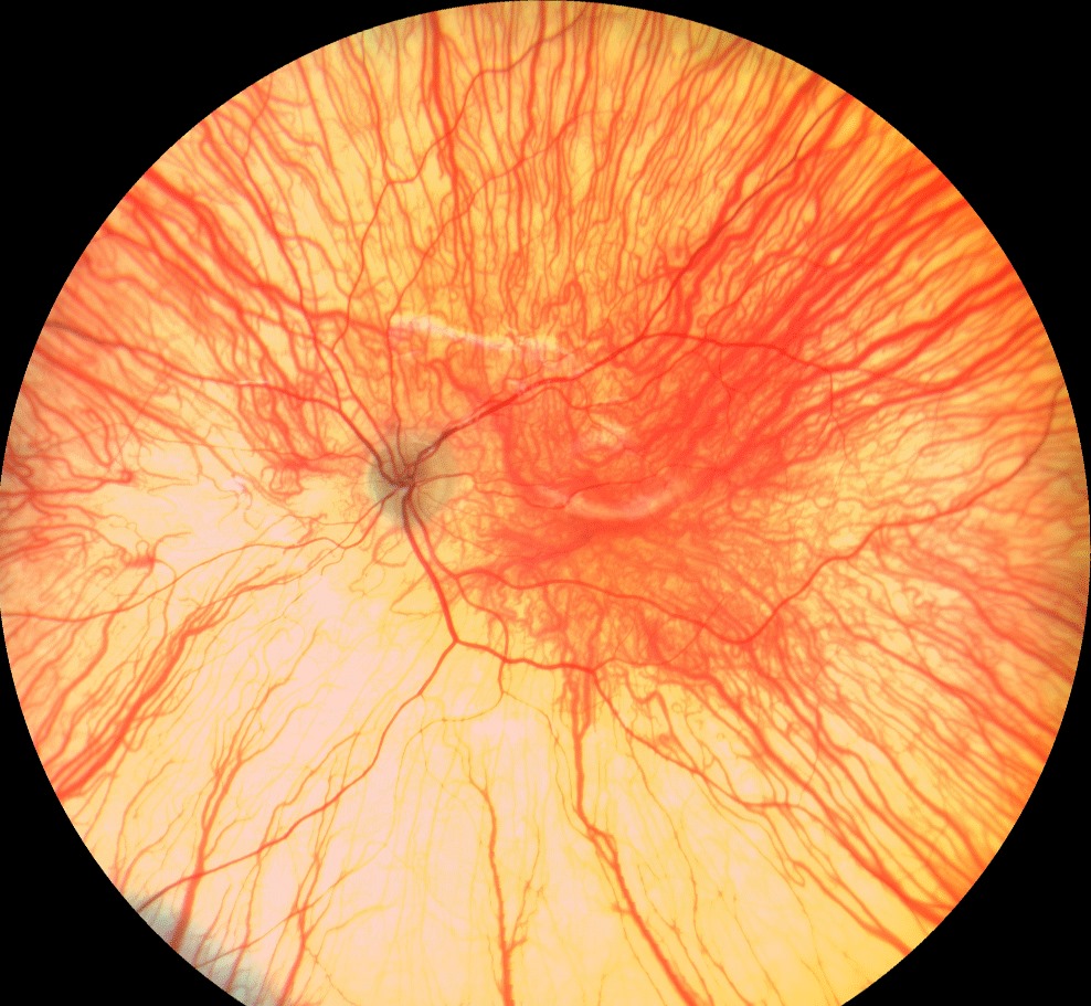
The choroid loses pigment, producing the orange-red sunset glow fundus.
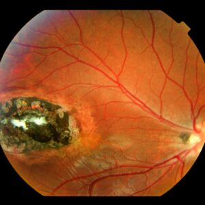
Small, round depigmented chorioretinal scars may also appear in the inferior periphery.
In addition, perilimbal vitiligo (Sugiura sign), poliosis, vitiligo and alopecia can develop.
Imaging Recovery in VKH Stages
Subretinal fluid resolves on OCT, and the choroid thins progressively.
For example, serial enhanced depth imaging can show subnormal choroidal thickness in long-standing disease.
Meanwhile, ICGA dark dots that return during the taper suggest ongoing choroidal activity.
Chronic Recurrent Phase
Recurrences usually present as granulomatous anterior uveitis.
Mutton-fat keratic precipitates, Koeppe and Busacca iris nodules and posterior synechiae are typical findings.
Posterior recurrences with fresh serous detachment are less common.
Therefore, a quiet posterior pole does not exclude active disease.
Complications of Late VKH Stages
Chronic inflammation leads to cataract, glaucoma, choroidal neovascularization and subretinal fibrosis.
Also, corticosteroids add to the cataract and pressure risk.
Glaucoma here needs combined control of pressure and inflammation (see uveitic glaucoma).
VKH Stages at a Glance
- Prodromal: headache, meningismus, tinnitus, scalp sensitivity; eye often normal
- Acute uveitic: disc hyperemia, serous detachments, septate fluid on OCT, pinpoint leaks on FA, shallow chamber
- Convalescent: sunset glow fundus, nummular scars, Sugiura sign, poliosis, thin choroid
- Chronic recurrent: granulomatous anterior uveitis, iris nodules, synechiae, cataract and glaucoma
Treatment Across VKH Stages
High-dose systemic corticosteroids are first-line in the acute uveitic phase.
They usually resolve serous detachments and restore vision.
However, a short course raises recurrence risk, so many experts advise a slow taper over at least six months.
Early immunomodulatory therapy may reduce progression to chronic disease, although most evidence is observational.
Finally, treatment cannot reverse established depigmentation or fibrosis.
Counseling Patients
Explain that the disease attacks pigment cells in the eye, skin and inner ear.
Then, warn that stopping steroids early invites relapse.
Also, prepare patients for possible whitening of hair, lashes and skin.
Follow-Up Through VKH Stages
Check acuity, pressure, anterior chamber cells and OCT at every visit during the taper.
In addition, repeat ICGA when relapse is suspected and the fundus looks quiet.
Pitfalls to Avoid
- Do not treat a shallow chamber in acute VKH with miotics
- Never stop corticosteroids abruptly once the detachments resolve
- Avoid labeling bilateral multifocal fluid as central serous chorioretinopathy
- Exclude sympathetic ophthalmia by asking about penetrating trauma or surgery


Document what you see
Two smartphone imaging tools built for everyday clinic use — one for the slit lamp, one for the fundus.
From Choroida — the team behind this siteReferences
- Moorthy RS, Inomata H, Rao NA. Vogt-Koyanagi-Harada syndrome. Surv Ophthalmol. 1995;39:265-292.
- Read RW, Holland GN, Rao NA, et al. Revised diagnostic criteria for Vogt-Koyanagi-Harada disease: report of an international committee on nomenclature. Am J Ophthalmol. 2001;131:647-652.
- Standardization of Uveitis Nomenclature (SUN) Working Group. Classification criteria for Vogt-Koyanagi-Harada disease. Am J Ophthalmol. 2021;228:205-211.
- Lavezzo MM, Sakata VM, Morita C, et al. Vogt-Koyanagi-Harada disease: review of a rare autoimmune disease targeting antigens of melanocytes. Orphanet J Rare Dis. 2016;11:29.
- American Academy of Ophthalmology. Basic and Clinical Science Course, Section 9: Uveitis and Ocular Inflammation. San Francisco: AAO; 2023.

Writes and reviews the clinical articles on Educate. Every post is checked against peer-reviewed sources before it goes live.