Wet vs dry AMD is the first distinction patients ask about.
Both are forms of age-related macular degeneration, but they behave very differently. Clear counseling reduces fear and improves follow-up.
What Is Dry AMD
Dry AMD involves drusen and gradual loss of retinal pigment epithelium and photoreceptors, and it accounts for most cases of AMD. Vision declines slowly over years, and therefore many patients notice little at first.
What Is Wet AMD
Wet AMD involves choroidal neovascularization, and the new vessels leak fluid and blood under the macula. Vision can fall within days or weeks, so wet disease is an emergency (see new treatments for neovascular AMD).
Fundus Explorer Pro
Photograph the retinal findings described here with the phone already in your pocket — 22 D optics and built-in illumination in one handheld unit.
From Choroida — the team behind this siteWet vs Dry AMD: Symptoms
Dry disease causes blur, trouble with dim light and slow central loss, while wet disease causes distortion, a dark spot or sudden blur. Straight lines look bent, so ask about new symptoms at every visit as well.
Wet vs Dry AMD on Examination
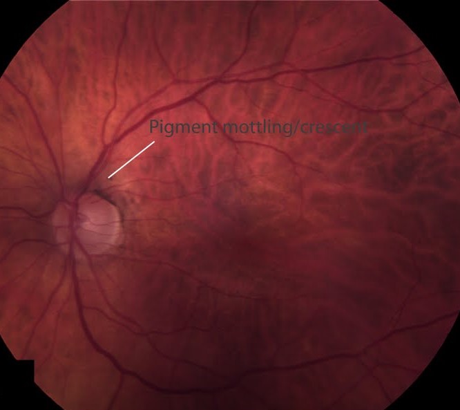
Dry AMD shows drusen and pigment changes, while geographic atrophy shows sharply outlined areas of loss. Wet AMD shows fluid, hemorrhage and exudate, so the fundus often gives the answer.
Imaging
OCT shows drusen, atrophy and fluid. Fluorescein angiography shows leakage. OCT angiography can show new vessels without dye. Therefore, image any patient with new symptoms.
Progression From Dry to Wet
A sizable minority of eyes with intermediate AMD progress to advanced disease within five years. Risk rises with large drusen, fellow-eye disease and smoking, so monitor high-risk eyes closely.
Home Monitoring
Patients can use an Amsler grid or a home device (see home monitoring devices for AMD). New distortion should prompt a same-week visit because early treatment preserves vision, and you should also remind patients to cover each eye separately.
Treatment of Wet AMD
Anti-VEGF injections are the standard. Ranibizumab, aflibercept, bevacizumab and faricimab all work. Regular follow-up prevents recurrence. Therefore, plan long-term care.
Treatment of Dry AMD
No treatment restores lost cells, but AREDS2 supplements slow progression in intermediate disease (see AREDS2 supplements). Complement inhibitors now slow geographic atrophy growth, so treatment options are growing.
Lifestyle Advice for Both Forms
Stop smoking, which is the strongest modifiable risk, and eat a diet rich in leafy vegetables and fish. Protect the eyes from strong sunlight, and also control blood pressure.
Low Vision Support
Many patients keep useful peripheral vision, and magnifiers, lighting and training help daily tasks (see low vision rehabilitation in AMD). Early referral improves independence, so do not wait for advanced loss.
Explaining Wet vs Dry AMD to Patients
Use a simple picture of the macula, describing dry AMD as a slow wearing down and wet AMD as leaking new vessels. Therefore, the urgency of wet disease becomes clear.
Wet vs Dry AMD: Practical Summary
Ask about distortion at every visit and check with OCT, and treat wet disease at once with anti-VEGF. Because dry disease can turn wet, teach home checks, and finally, support lifestyle change.
Pitfalls to Avoid
- Do not tell patients that dry AMD can never turn wet
- Never delay injections in active wet disease
- Avoid ignoring new distortion
- Do not skip smoking cessation advice
Wet vs Dry AMD in the Fellow Eye
Patients with wet AMD in one eye face higher risk in the other, so monitor the fellow eye closely. Because early detection matters, teach home checks, and therefore schedule frequent visits.
Wet vs Dry AMD and Supplements
AREDS2 supplements help intermediate dry AMD, but they do not treat wet disease directly. Because supplements vary, recommend the studied formula, and then explain realistic benefits.
Wet vs Dry AMD and Geographic Atrophy
Geographic atrophy is the advanced dry form, and complement inhibitors slow the growth of atrophy. Because treatment needs injections, discuss burdens, so decide together with the patient.
Wet vs Dry AMD and Driving
Central vision loss affects reading and driving, so test visual acuity against legal standards. If vision falls, advise on safe alternatives, and in addition, refer for low vision services.
Wet vs Dry AMD and Anxiety
Fear of blindness is common in AMD, and honest, hopeful information eases worry. Because most patients keep useful vision, say so, and then offer support groups.
Wet vs Dry AMD: Frequently Asked Questions
Patients ask whether diet can reverse AMD, and it cannot, but good habits slow progression. They ask whether eye injections hurt, and most feel only pressure, so reassure them.
Wet vs Dry AMD: Practical Summary
If the patient reports new distortion, image the macula at once, and because wet disease needs fast injections, book the visit within days. However, reassure patients with dry disease that slow loss is usual, and finally, repeat the education at every visit.


Document what you see
Two smartphone imaging tools built for everyday clinic use — one for the slit lamp, one for the fundus.
From Choroida — the team behind this siteReferences
- Mitchell P, Liew G, Gopinath B, Wong TY. Age-related macular degeneration. Lancet. 2018;392:1147-1159.
- Ferris FL 3rd, Wilkinson CP, Bird A, et al. Clinical classification of age-related macular degeneration. Ophthalmology. 2013;120:844-851.
- Age-Related Eye Disease Study Research Group. A randomized, placebo-controlled, clinical trial of high-dose supplementation with vitamins C and E, beta carotene, and zinc for age-related macular degeneration and vision loss: AREDS report no. 8. Arch Ophthalmol. 2001;119:1417-1436.
- CATT Research Group. Ranibizumab and bevacizumab for neovascular age-related macular degeneration. N Engl J Med. 2011;364:1897-1908.
Test yourself
A few questions straight from this article.
-
Which process defines dry age-related macular degeneration?
Dry AMD involves drusen and gradual loss of retinal pigment epithelium and photoreceptors, with vision declining slowly over years. -
What is the underlying lesion in wet age-related macular degeneration?
Wet AMD involves choroidal neovascularization; the new vessels leak fluid and blood under the macula, so vision can fall within days or weeks. -
Which symptom points to wet rather than dry age-related macular degeneration?
Dry disease causes blur, dim-light difficulty and slow central loss, while wet disease causes distortion, a dark spot or sudden blur, with straight lines looking bent. -
How does geographic atrophy appear on fundus examination in advanced dry AMD?
Dry AMD shows drusen and pigment changes, geographic atrophy shows sharply outlined areas of loss, and wet AMD shows fluid, hemorrhage and exudate. -
Which imaging test can show the new vessels of wet AMD without injecting dye?
OCT shows drusen, atrophy and fluid, fluorescein angiography shows leakage, and OCT angiography can show new vessels without dye. -
Which features raise the risk of intermediate AMD progressing to advanced disease?
Risk of progression rises with large drusen and fellow-eye disease, and smoking raises it further, so high-risk eyes are monitored closely. -
What is the standard treatment for wet age-related macular degeneration?
Anti-VEGF injections are the standard; ranibizumab, aflibercept, bevacizumab and faricimab all work, and long-term follow-up is planned. -
In which stage of dry AMD do AREDS2 supplements slow progression?
AREDS2 supplements slow progression in intermediate dry AMD but do not treat wet disease directly, and no treatment restores lost cells. -
Which treatment now slows the growth of geographic atrophy?
Geographic atrophy is the advanced dry form, and complement inhibitors slow its growth; because they need injections, burdens are discussed with the patient. -
What is the strongest modifiable risk factor for age-related macular degeneration?
Stopping smoking addresses the strongest modifiable risk; a diet rich in leafy vegetables and fish and blood pressure control are also advised.

Writes and reviews the clinical articles on Educate. Every post is checked against peer-reviewed sources before it goes live.