Toxoplasma vs toxocara is a common differential when a clinician finds a focal chorioretinal lesion with vitreous inflammation.
Both parasites reach the eye, but they behave very differently at the fundus.
As a result, age, vitritis, lesion shape, serology and treatment usually separate them quickly.
Two Parasites, Two Life Cycles
Toxoplasma gondii is an intracellular protozoan, and cats are its definitive host.
Toxocara canis and Toxocara cati, in contrast, are roundworms of dogs and cats.
Children ingest the eggs from soil, and a single larva can migrate to the eye.
Age in Toxoplasma vs Toxocara
Ocular toxocariasis typically affects children and young adults.
Most series describe a child with unilateral vision loss, strabismus or leukocoria.
Toxoplasmosis, however, appears at any age.
Recurrent attacks often present in young adults.
Fundus Explorer Pro
Photograph the retinal findings described here with the phone already in your pocket — 22 D optics and built-in illumination in one handheld unit.
From Choroida — the team behind this siteLaterality and History
Toxocara disease is almost always unilateral.
Therefore, a history of puppy contact, pica or play in soil supports the diagnosis.
Toxoplasmosis is also usually unilateral, although congenital infection may leave scars in both eyes.
Vitritis in Toxoplasma vs Toxocara
Active toxoplasma retinochoroiditis produces dense vitritis over a white focus.
The result is the classic “headlight in the fog” appearance.
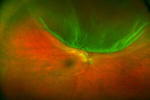
Peripheral and posterior toxocara granulomas usually show a quieter vitreous.
The chronic endophthalmitis form of toxocariasis, however, can cause marked vitreous haze.
Lesion Type in Toxoplasma vs Toxocara
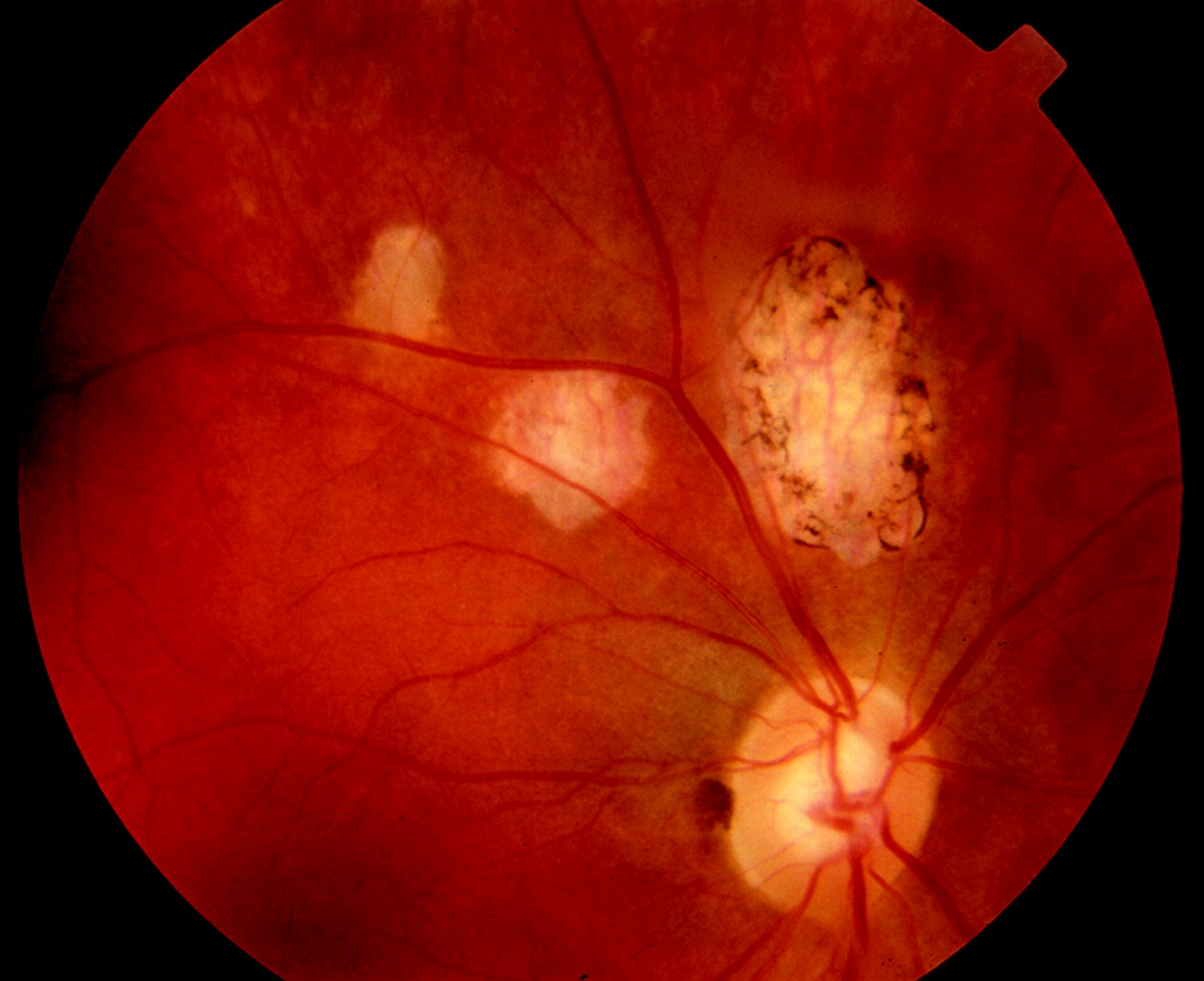
Toxoplasma causes a fluffy, full-thickness necrotizing retinitis with fuzzy margins.
The new focus typically sits beside a pigmented scar, which suggests reactivation.
Toxocara, on the other hand, produces a firm, elevated white granuloma with sharper borders.
Also, it often anchors vitreous bands or a retinal fold.
Three Toxocara Patterns
Toxocariasis typically follows one of three clinical patterns.
- First, a posterior pole granuloma, often near the macula or optic disc
- Next, a peripheral granuloma with a fibrous band or fold toward the disc
- Finally, chronic endophthalmitis with dense vitritis and possible leukocoria
Scars and Recurrence in Toxoplasma vs Toxocara
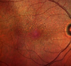
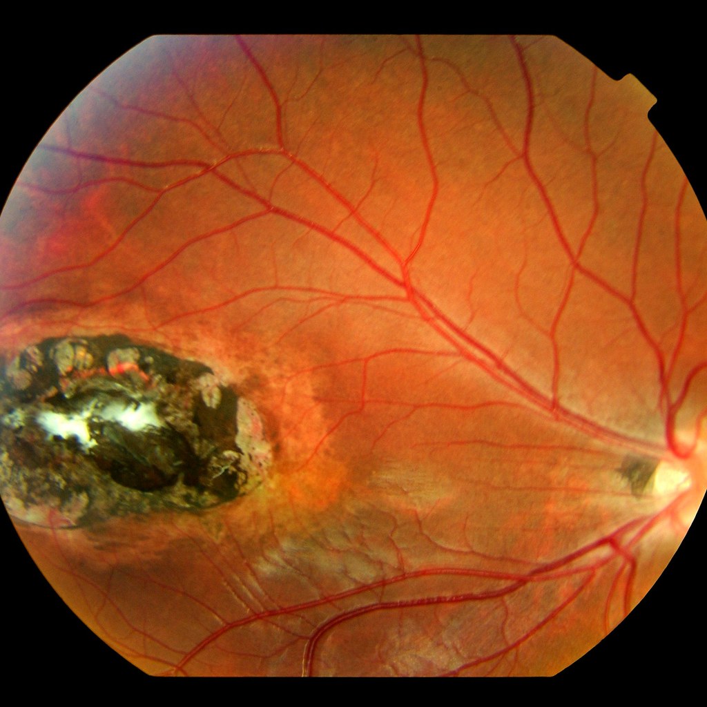
Toxoplasma scars are atrophic and punched out, with dense pigment at the edge.
Moreover, recurrences tend to occur at the scar border (see reactivation signs at old scars).
Instead, toxocara vision loss comes from traction, macular involvement or retinal detachment (see parasitic endophthalmitis).
Serology in Toxoplasma vs Toxocara
Serum toxoplasma IgG is common in the general population.
Therefore, a positive result supports the diagnosis but cannot prove it.
A negative IgG, however, makes ocular toxoplasmosis unlikely in an immunocompetent patient.
Toxocara ELISA titers may be low or negative in ocular disease.
As a result, a low titer does not exclude toxocariasis when the fundus fits.
Intraocular Fluid Testing
Aqueous humor testing helps in atypical cases.
For example, PCR for Toxoplasma DNA and the Goldmann-Witmer coefficient can confirm local infection.
Intraocular toxocara antibody testing can also support the diagnosis when serum titers are low.
Imaging Clues in Toxoplasma vs Toxocara
OCT over active toxoplasmosis shows thickened, hyperreflective inner retina with overlying vitreous cells.
Later, the healed area shows retinal thinning and atrophy.
Toxocara granulomas appear on OCT as elevated hyperreflective masses with traction on the nearby retina.
In addition, B-scan ultrasonography helps in children when media opacity hides the fundus.
Ruling Out Retinoblastoma
A child with leukocoria and a white mass needs retinoblastoma excluded first.
Toxocara granulomas usually lack calcification on ultrasound, while retinoblastoma often contains calcium.
Therefore, refer any doubtful case to an ocular oncologist before treating inflammation.
Treating Ocular Toxoplasmosis
Many small peripheral lesions in immunocompetent patients heal without therapy.
However, treat lesions near the macula or disc, large lesions and severe vitritis.
Classic therapy combines pyrimethamine, sulfadiazine and folinic acid, often with oral corticosteroids.
Trimethoprim-sulfamethoxazole and intravitreal clindamycin with dexamethasone are accepted alternatives in many centers.
Treatment shortens the active phase, but evidence that it improves final vision remains limited.
Treating Ocular Toxocariasis
Corticosteroids control the inflammation around the dying larva.
The role of albendazole remains uncertain, and practice varies between specialists.
Vitrectomy addresses traction, epiretinal membranes and retinal detachment.
Visual outcome depends mainly on macular involvement and traction.
Steroid Safety in Toxoplasma vs Toxocara
Steroids alone can worsen toxoplasma retinochoroiditis.
Therefore, never give periocular or systemic steroids without antiparasitic cover.
Clinicians often treat toxocara inflammation with steroids, with or without albendazole.
Counseling the Patient
Explain that toxoplasma scars may reactivate, often years later.
Ask patients to return quickly if floaters or blurred vision appear.
For toxocariasis, advise deworming of pets and hand washing after soil contact.
Parents also need to know that the child’s vision loss may be permanent.
Pitfalls to Avoid
- Do not assume a white lesion in a child is toxocara before excluding retinoblastoma
- Never treat suspected toxoplasmosis with steroids alone
- Do not rely on serum toxocara titers to exclude the disease
- Avoid missing atypical toxoplasmosis in elderly or immunocompromised patients


Document what you see
Two smartphone imaging tools built for everyday clinic use — one for the slit lamp, one for the fundus.
From Choroida — the team behind this siteReferences
- Holland GN. Ocular toxoplasmosis: a global reassessment. Part I: epidemiology and course of disease. Am J Ophthalmol. 2003;136:973-988.
- Soheilian M, Ramezani A, Azimzadeh A, et al. Randomized trial of intravitreal clindamycin and dexamethasone versus pyrimethamine, sulfadiazine, and prednisolone in treatment of ocular toxoplasmosis. Ophthalmology. 2011;118:134-141.
- Shields JA. Ocular toxocariasis. A review. Surv Ophthalmol. 1984;28:361-381.
- Woodhall D, Starr MC, Montgomery SP, et al. Ocular toxocariasis: epidemiologic, anatomic, and therapeutic variations based on a survey of ophthalmic subspecialists. Ophthalmology. 2012;119:1211-1217.

Writes and reviews the clinical articles on Educate. Every post is checked against peer-reviewed sources before it goes live.