
A sunset glow fundus is the name for a fundus that has turned uniformly orange to red because the pigment in the choroid has been lost.
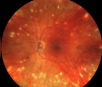
The retina is clear, the retinal pigment epithelium is relatively intact, and the choroidal vessels are easily visible through a pale background.
The result is a glowing, sunset-colored posterior pole.
How it develops
In Vogt-Koyanagi-Harada disease, the immune attack is directed at melanocytes.
During the acute phase, the choroid is thickened and inflamed, and there are serous retinal detachments.
After the acute phase, melanocytes are destroyed and the choroid loses pigment.
Months later, in the convalescent stage, the fundus appears orange-red, often with scattered pigment clumps and depigmented scars at the RPE level (see VKH disease).
Other features of the convalescent stage
- Sugiura sign, a perilimbal vitiligo, seen in a large proportion of patients
- Poliosis of lashes or eyebrows, vitiligo, and alopecia
- Dalen-Fuchs-like nodules scarring into small pigmented spots (see Dalen-Fuchs nodules)
- Peripapillary atrophy
- Cataract, glaucoma, and subretinal fibrosis as complications
Fundus Explorer Pro
Photograph the retinal findings described here with the phone already in your pocket — 22 D optics and built-in illumination in one handheld unit.
From Choroida — the team behind this siteWhy it matters clinically
The sign is not an indicator of active disease.
It documents past, often unrecognized, inflammation.
In a young patient presenting for the first time with orange-red fundus and no history, VKH should be considered, and a careful history asking about previous headache, tinnitus, hearing change, or neck stiffness may reveal it.
The sign also warns about the risk of chronic recurrent granulomatous uveitis, which occurs in many patients who were undertreated at the start of the illness.
Other conditions that cause a similar appearance
Choroidal depigmentation can occur in sympathetic ophthalmia, in some forms of albinism, and in pale fundi of fair-skinned patients.
The key differences are that VKH and sympathetic ophthalmia are acquired, bilateral, and accompanied by a history of inflammation, whereas albinism is congenital and associated with nystagmus and foveal hypoplasia.
Imaging and monitoring
- Fundus autofluorescence shows patchy loss of signal where the RPE has been damaged
- OCT will show a thin choroid in the late stage, in contrast with the thick choroid of the acute phase
- Indocyanine green angiography may reveal persistent choroidal inflammation even when the clinical picture looks quiet, and some centers use it to monitor subclinical activity
Staging VKH from the fundus
The disease passes through four phases, and the fundus changes with each.
In the prodromal phase, which lasts a few days, there are neurological symptoms such as headache, tinnitus and meningism, and the fundus may be normal.
In the acute uveitic phase, over the next several weeks, there is diffuse choroiditis, disc hyperemia and multifocal serous detachments.
The convalescent phase follows, with depigmentation of the choroid and the appearance of the sunset glow fundus, and the chronic recurrent phase is characterized by recurrent granulomatous anterior uveitis and complications.
An examiner who understands the sequence can usually place a patient in the correct phase from the fundus alone.
A distinctive point is that depigmentation also happens in the skin and hair, and the timing differs.
Poliosis, vitiligo and alopecia typically show up weeks to months after the eye disease begins, so a patient who is seen early may not yet have them.
Management
Treatment is directed at preventing recurrences and at managing complications.
Early, aggressive and prolonged immunosuppression in the acute phase, usually high-dose corticosteroids followed by a long taper with or without steroid-sparing agents, reduces the chance of progression to sunset glow fundus and chronic disease.
A note on the differential diagnosis
In a patient who presents late with an orange fundus and recurrent anterior uveitis, the main alternatives are sympathetic ophthalmia and, less often, tuberculosis or syphilis, which can both produce choroidal depigmentation. A history of previous penetrating injury or surgery, and the presence of extraocular signs such as poliosis, vitiligo, and hearing loss, usually decide between them. When the history is unclear, serologic tests for syphilis and tuberculosis screening are reasonable before long-term immunosuppression is started.
Long-term care
Patients with a sunset glow fundus remain at risk for glaucoma, cataract, and choroidal neovascularization, so regular examination is important. The treating team should watch for early recurrence of anterior inflammation, which is a signal to reassess the immunosuppressive plan.
Documenting the stage
Record the stage of disease, the extent of depigmentation, and the associated features such as Sugiura sign and poliosis, along with the treatments given. Photographs of the fundus and of the limbus provide a useful record. A clear staging note helps other clinicians, because patients often move between clinics over the years.
Prognosis
With timely and adequate therapy, many patients retain good vision despite the fundus change.
Patients who progress to the chronic recurrent stage need long-term follow-up for glaucoma, cataract, and choroidal neovascularization.


Document what you see
Two smartphone imaging tools built for everyday clinic use — one for the slit lamp, one for the fundus.
From Choroida — the team behind this siteReferences
- Read RW, Holland GN, Rao NA, et al. Revised diagnostic criteria for Vogt-Koyanagi-Harada disease. Am J Ophthalmol. 2001;131:647-652.
- Moorthy RS, Inomata H, Rao NA. Vogt-Koyanagi-Harada syndrome. Surv Ophthalmol. 1995;39:265-292.
- Herbort CP Jr, Abu El Asrar AM, Yamamoto JH, et al. Reappraisal of the management of Vogt-Koyanagi-Harada disease: sunset glow fundus is no more a fatality. Int Ophthalmol. 2017;37:1383-1395.
- Rao NA. Pathology of Vogt-Koyanagi-Harada disease. Int Ophthalmol. 2007;27:81-85.
Test yourself
A few questions straight from this article.
-
What gives the fundus its uniform orange-red colour in a sunset glow fundus?
Choroidal pigment is lost while the retina stays clear and the RPE remains relatively intact, so choroidal vessels show through a pale background as a glowing orange-red pole. -
In Vogt-Koyanagi-Harada disease, the immune attack is directed against which cell type?
The immune attack in VKH targets melanocytes; their destruction after the acute phase leaves the choroid depigmented and produces the sunset glow appearance. -
What is the Sugiura sign seen in convalescent Vogt-Koyanagi-Harada disease?
The Sugiura sign is a perilimbal vitiligo, one of the extraocular depigmentation features of the convalescent stage of VKH. -
What does a sunset glow fundus indicate about disease activity in Vogt-Koyanagi-Harada disease?
The sign is not an indicator of active disease; it records previous and often unrecognised inflammation, and warns of the risk of chronic recurrent granulomatous uveitis. -
Which systemic symptoms should be sought when a young patient presents with an orange-red fundus and no known history?
A careful history asking about previous headache, tinnitus, hearing change or neck stiffness may reveal unrecognised VKH behind an orange-red fundus. -
Which features distinguish albinism from Vogt-Koyanagi-Harada disease as a cause of choroidal depigmentation?
Albinism is congenital and associated with nystagmus and foveal hypoplasia, whereas VKH and sympathetic ophthalmia are acquired, bilateral and accompanied by a history of inflammation. -
What does OCT show of the choroid in the late stage of Vogt-Koyanagi-Harada disease?
OCT shows a thin choroid in the late stage, contrasting with the thickened choroid that characterises the acute uveitic phase. -
What may indocyanine green angiography reveal in Vogt-Koyanagi-Harada disease that looks clinically quiet?
ICG angiography may reveal persistent choroidal inflammation even when the clinical picture looks quiet, and some centres use it to monitor subclinical activity. -
When do poliosis, vitiligo and alopecia typically appear relative to the onset of eye disease in Vogt-Koyanagi-Harada disease?
Skin and hair depigmentation lags behind the eye disease, typically appearing weeks to months after onset, so a patient seen early may not yet show them. -
Which treatment approach reduces the chance of progression to sunset glow fundus and chronic disease?
Early, aggressive and prolonged immunosuppression during the acute phase, usually high-dose corticosteroids with a long taper, reduces progression to sunset glow fundus and chronic disease.
