Tear meniscus height is the vertical depth of the tear strip along the lower lid margin.
It offers a quick, noninvasive estimate of aqueous tear volume.
Therefore, it complements Schirmer testing and tear break-up time in the dry eye workup.
What the Tear Meniscus Is
Each lid margin holds a concave strip of tear fluid.
Together, the upper and lower menisci hold most of the tear volume.
As a result, their size reflects aqueous output from the lacrimal glands.
Clinicians usually measure the lower meniscus because it is easier to see.
Why Tear Meniscus Height Matters
Tear secretion falls in aqueous deficiency, and the meniscus shrinks.
A thin meniscus also supplies less fluid to resurface the cornea with each blink.
In addition, the test takes seconds and needs no contact with the eye.
All-fit Slit-Lamp Adapter
Record and share exactly what you see at the slit lamp. One adapter fits any slit lamp or surgical microscope — and any smartphone.
From Choroida — the team behind this siteSlit-Lamp Measurement of Tear Meniscus
First, center a narrow vertical beam on the lower lid below the pupil.
Next, compare the tear strip with the known beam height or an eyepiece graticule.
Keep the light dim to limit reflex tearing (see slit lamp illumination).
Then record the height in millimeters for each eye.
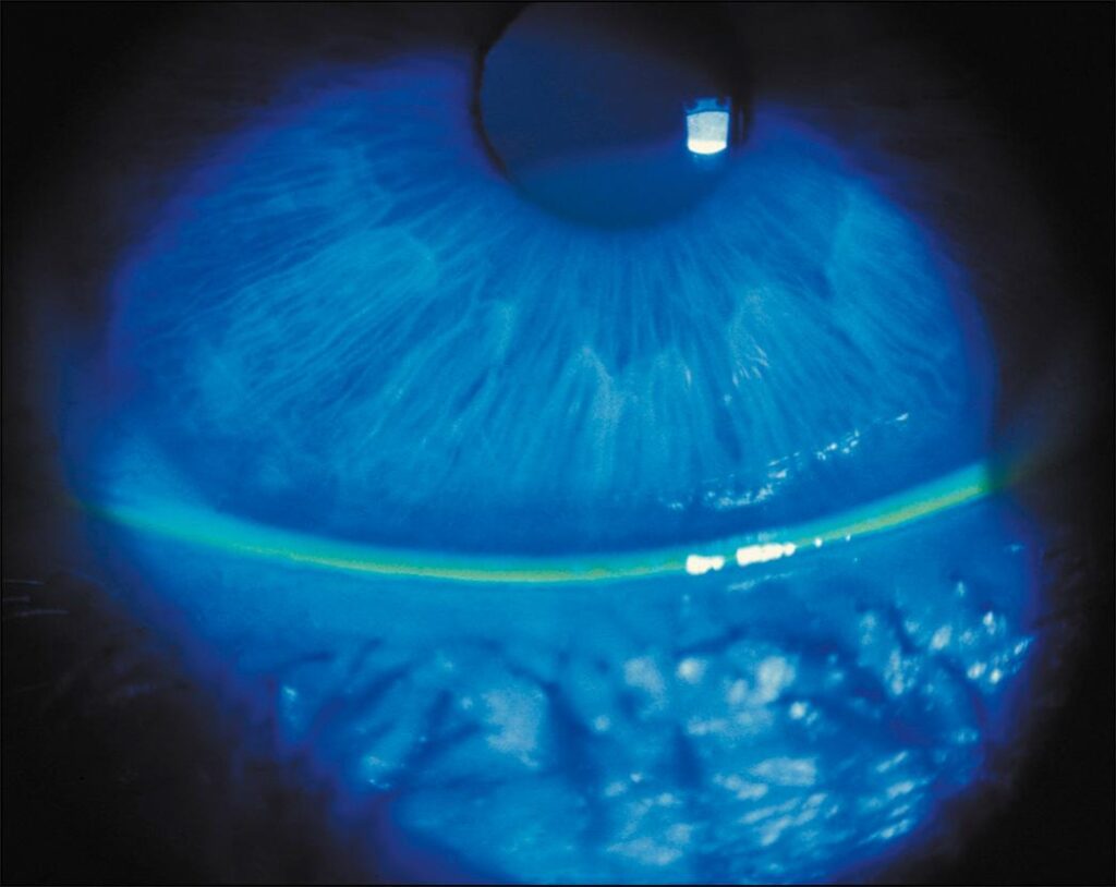
Fluorescein and the Tear Lake
Fluorescein makes the meniscus easy to see under cobalt blue light.
However, the drop adds volume and may provoke tearing.
Therefore, measure before dye, or use a lightly moistened strip.
Then wait a few blinks before reading the height.
Normal Tear Meniscus Values
Published normal values vary with method, age and population.
Many clinicians regard a lower meniscus below about 0.2 mm as reduced.
However, cutoffs from one technique or device do not transfer directly to another.
Age, lid position and environment also shift the baseline.
OCT Imaging of the Tear Meniscus
Anterior segment OCT shows the meniscus in cross section without touching the eye (see anterior segment OCT).
A vertical scan crosses the central lower lid.
Then calipers or software measure height, depth and cross-sectional area.
Because OCT needs no dye, it disturbs the tears less.
OCT Parameters and Repeatability
OCT meniscus height and area correlate with Schirmer results and symptoms in several studies.
Also, OCT readings repeat better than subjective slit-lamp estimates.
However, the meniscus changes after each blink, so scan timing matters.
Many protocols capture the image a few seconds after a blink.
Aqueous Deficiency and Tear Meniscus
A low meniscus supports aqueous-deficient dry eye, including Sjögren disease.
Schirmer values are usually low as well.
Also, look for interpalpebral staining and corneal filaments.
Then consider a systemic workup when the pattern fits.
Evaporative Dry Eye and Tear Meniscus
In evaporative dry eye, lacrimal secretion is often adequate.
As a result, the meniscus may look normal while the film breaks up quickly.
Meibomian gland dysfunction is the usual driver.
Therefore, pair meniscus height with tear break-up time and lid margin assessment.
Mixed Disease
Many patients have both mechanisms.
For example, a low meniscus with short break-up time suggests mixed disease.
This pattern is common in older adults with chronic lid disease.
Then treatment should address both lubrication and the lids.
A High Tear Meniscus
A tall meniscus does not always mean healthy tears.
Nasolacrimal obstruction raises the tear lake and causes epiphora.
Reflex tearing from irritation can also inflate it briefly.
Therefore, assess lacrimal drainage when a watery patient shows a high meniscus.
Lid and Conjunctival Factors
Conjunctivochalasis folds can interrupt the lower meniscus.
In addition, lid notching or ectropion distorts its shape.
Measure away from these areas, or note the reading as unreliable.
Contact lens wear can also alter readings.
Limits of the Tear Meniscus Test
The meniscus reflects tear volume at a single moment.
However, volume shifts with time of day, humidity and blink rate.
Slit-lamp grading is subjective and varies between observers.
Therefore, no single reading should confirm or exclude dry eye.
Tear Meniscus in Follow-Up
Serial measurements can track response to treatment, such as punctal occlusion.
For example, plugs may raise meniscus height when aqueous deficiency drives the disease.
Use the same method, eye and time of day each visit.
Also, document whether fluorescein was instilled.
Explaining the Result to Patients
Patients readily understand the tear lake as a reservoir along the lid.
A low reservoir helps explain burning and fluctuating blur.
Then a slit-lamp photo can show why lubrication or plugs are advised.
However, avoid presenting one number as a full diagnosis.
Pitfalls to Avoid
- Measuring after a large fluorescein drop
- Using a bright beam that triggers reflex tearing
- Reading over conjunctivochalasis folds
- Applying OCT cutoffs from another device
- Diagnosing dry eye from the meniscus alone
- Missing nasolacrimal obstruction behind a high meniscus


Document what you see
Two smartphone imaging tools built for everyday clinic use — one for the slit lamp, one for the fundus.
From Choroida — the team behind this siteReferences
- Wolffsohn JS, Arita R, Chalmers R, et al. TFOS DEWS II diagnostic methodology report. Ocul Surf. 2017;15:539-574.
- Craig JP, Nichols KK, Akpek EK, et al. TFOS DEWS II definition and classification report. Ocul Surf. 2017;15:276-283.
- Mainstone JC, Bruce AS, Golding TR. Tear meniscus measurement in the diagnosis of dry eye. Curr Eye Res. 1996;15:653-661.
- Tung CI, Perin AF, Gumus K, Pflugfelder SC. Tear meniscus dimensions in tear dysfunction and their correlation with clinical parameters. Am J Ophthalmol. 2014;157:301-310.
- Shen M, Li J, Wang J, et al. Upper and lower tear menisci in the diagnosis of dry eye. Invest Ophthalmol Vis Sci. 2009;50:2722-2726.

Writes and reviews the clinical articles on Educate. Every post is checked against peer-reviewed sources before it goes live.