Optic neuritis mimics are optic neuropathies that resemble demyelinating optic neuritis at presentation.
They include ischemic, compressive, infiltrative, hereditary and inflammatory disorders.
Therefore, every new case needs a deliberate search for features that do not fit.
Why Optic Neuritis Mimics Matter
Stunkel and colleagues studied patients referred to a neuro-ophthalmology clinic with presumed optic neuritis.
Many of them turned out to have another diagnosis.
As a result, some received unnecessary steroids while the true cause went untreated.
The Typical Demyelinating Profile
Classic optic neuritis affects young adults, more often women, with pain on eye movement.
Vision falls over hours to days, and a relative afferent pupillary defect appears in unilateral cases.
In addition, about two thirds of eyes in the ONTT had a normal disc.
Most patients then start to improve within three weeks, even without treatment.
Fundus Explorer Pro
Photograph the retinal findings described here with the phone already in your pocket — 22 D optics and built-in illumination in one handheld unit.
From Choroida — the team behind this siteRed Flags for Optic Neuritis Mimics
- Age over 50 or vascular risk factors
- Painless visual loss
- Progression beyond two weeks
- No recovery after one month
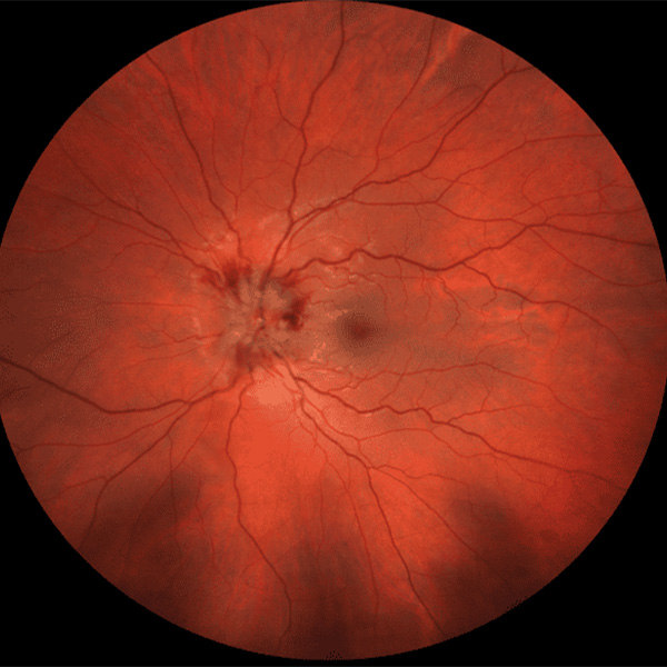
- Severe disc edema with hemorrhages or exudates
- Simultaneous bilateral involvement
- Headache, jaw claudication or weight loss
Anterior Ischemic Optic Neuropathy
Ischemic optic neuropathy typically causes sudden, painless loss, often noticed on waking.
The disc is swollen at onset, often sectorally, with flame hemorrhages.
In addition, the fellow eye usually shows a small, crowded disc.
Recovery is limited, while optic neuritis tends to improve (see anterior ischemic optic neuropathy).
Giant Cell Arteritis
Any patient over 50 with disc swelling needs urgent exclusion of giant cell arteritis.
Ask about headache, scalp tenderness and jaw claudication, then check ESR, CRP and platelets that day.
Because the fellow eye is at risk, start high-dose steroids before biopsy when suspicion is high.
Compressive Lesions in Optic Neuritis Mimics
Meningioma, pituitary adenoma and orbital masses compress the nerve slowly and usually painlessly.
However, patients may report sudden loss they noticed only when covering one eye.
In addition, proptosis, optociliary shunt vessels or a junctional field defect point to compression.
Infiltrative and Inflammatory Causes
Sarcoidosis, lymphoma, leukemia and syphilis can infiltrate the optic nerve.
Therefore, look carefully for anterior chamber cells, vitritis and vasculitis at the slit lamp.
Relapse whenever steroids are tapered should also raise suspicion of sarcoidosis.
Leber Hereditary Optic Neuropathy
LHON typically causes painless, severe central loss in young men.
The second eye usually follows within weeks to months.
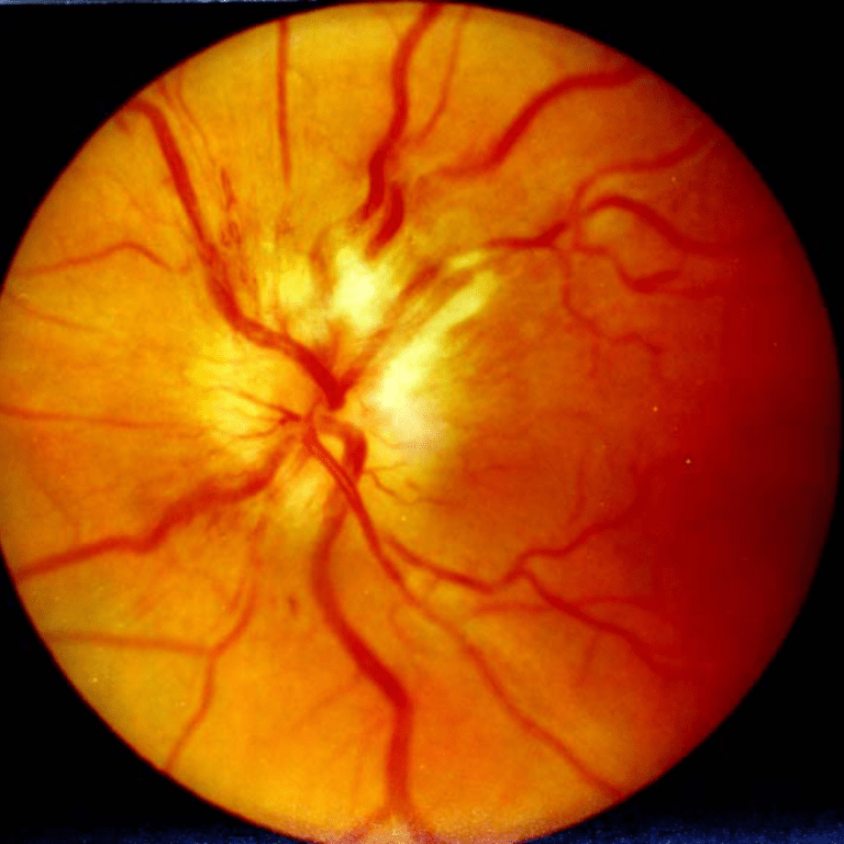
The fundus shows disc hyperemia and peripapillary telangiectasia, yet the disc does not leak on angiography.
Then mitochondrial genetic testing confirms it (see LHON).
Optic Perineuritis
Optic perineuritis inflames the nerve sheath, often in older patients with prominent pain.
Central vision may be relatively spared, while MRI shows enhancement around the nerve.
It responds quickly to steroids.
However, it may relapse after rapid tapering.
Toxic and Nutritional Causes
Toxic and nutritional optic neuropathies cause bilateral, symmetric, painless loss with cecocentral scotomas.
Therefore, ask about ethambutol, methanol, alcohol and restrictive diets.
Retinal Optic Neuritis Mimics
Some outer retinal disorders reduce vision with a nearly normal fundus.
For example, MEWDS and acute zonal occult outer retinopathy may enlarge the blind spot.
OCT then shows ellipsoid zone loss, which places the problem in the retina.
Neuroretinitis, with disc edema and a macular star, also calls for Bartonella and syphilis testing.
Exam Clues That Separate Optic Neuritis Mimics
Color loss out of proportion to acuity favors optic nerve disease.
In contrast, retinal disease usually spares the pupil reactions.
Also look for a crowded disc, early pallor or vitreous cells.
Imaging Optic Neuritis Mimics
Contrast MRI of the orbits and brain is the main test (see what the MRI predicts).
In contrast, long-segment or chiasmal enhancement suggests aquaporin-4 or MOG antibody disease.
Next, look for sheath enhancement, masses and adjacent sinus disease.
Blood Tests for Optic Neuritis Mimics
- ESR, CRP and platelets in patients over 50
- Aquaporin-4 and MOG antibodies in severe or bilateral cases
- Syphilis serology in atypical cases
- ACE and chest imaging for suspected sarcoidosis
- Vitamin B12, folate and copper in symmetric loss
Steroids and Optic Neuritis Mimics
In the ONTT, intravenous steroids sped recovery but did not change final vision.
Therefore, a good response proves little, because lymphoma and sarcoidosis can also improve.
A patient without recovery at one month therefore needs the diagnosis reconsidered.
Explain this uncertainty to patients before starting treatment.
Pitfalls to Avoid
- Do not label painless loss in an older patient as optic neuritis
- Never delay ESR and CRP because arteritis can blind the fellow eye
- Avoid stopping the workup after a normal brain MRI
- Remember that a normal disc does not exclude compression


Document what you see
Two smartphone imaging tools built for everyday clinic use — one for the slit lamp, one for the fundus.
From Choroida — the team behind this siteReferences
- Optic Neuritis Study Group. The clinical profile of optic neuritis: experience of the Optic Neuritis Treatment Trial. Arch Ophthalmol. 1991;109:1673-1678.
- Beck RW, Cleary PA, Anderson MM Jr, et al. A randomized, controlled trial of corticosteroids in the treatment of acute optic neuritis. N Engl J Med. 1992;326:581-588.
- Stunkel L, Kung NH, Wilson B, McClelland CM, Van Stavern GP. Incidence and causes of overdiagnosis of optic neuritis. JAMA Ophthalmol. 2018;136:76-81.
- Petzold A, Fraser CL, Abegg M, et al. Diagnosis and classification of optic neuritis. Lancet Neurol. 2022;21:1120-1134.
- Purvin V, Kawasaki A, Jacobson DM. Optic perineuritis: clinical and radiographic features. Arch Ophthalmol. 2001;119:1299-1306.

Writes and reviews the clinical articles on Educate. Every post is checked against peer-reviewed sources before it goes live.