Coats disease stages follow the Shields classification, which grades the disease by telangiectasia, exudation, detachment and secondary complications.
The system links fundus findings to treatment choice and visual prognosis.
Therefore, clinicians can use one examination to plan care and counsel families.
What Coats Disease Stages Describe
Coats disease is an idiopathic retinal vascular disorder that typically affects one eye in young boys.
Abnormal vessels leak lipid-rich fluid into and under the retina.
For a general overview, see Coats disease.
Examining the Eye Before Staging
Young children often present with leukocoria, strabismus or reduced vision.
First, perform a dilated examination with scleral depression, under anesthesia when necessary.
Next, map the telangiectasia, exudation and any subretinal fluid.
Fundus Explorer Pro
Photograph the retinal findings described here with the phone already in your pocket — 22 D optics and built-in illumination in one handheld unit.
From Choroida — the team behind this siteStage 1: Telangiectasia Only
The examiner sees telangiectatic vessels without visible exudation.
Vision usually remains good because the macula stays uninvolved.
Observation may suit a nonleaking lesion, while laser can treat leaking vessels.
Stage 2: Exudation
In stage 2A, yellow exudate spares the fovea, and laser or cryotherapy treats the leaking vessels.
In stage 2B, exudate involves the fovea, and central vision often falls.
Treatment closes the vessels, but it cannot reliably restore foveal function after lipid and fibrosis damage the photoreceptors.
A dense foveal nodule therefore suggests a guarded outcome.
Stage 3: Retinal Detachment
Stage 3A is a subtotal exudative detachment, with the fovea spared in 3A1 and involved in 3A2.
Cryotherapy often suits elevated retina better than laser.
Stage 3B is total detachment, so ablation alone may fail because fluid blocks the treatment effect.
Surgeons may then combine fluid drainage, cryotherapy and vitreoretinal procedures.
Stage 4: Total Detachment and Glaucoma
Stage 4 adds secondary glaucoma to total retinal detachment.
Neovascular glaucoma can cause pain, corneal edema and high pressure.
Topical medication may provide comfort, while cyclodestructive treatment may help selected eyes.
However, enucleation often becomes the most reliable option for a blind, painful eye.
Stage 5: Advanced End-Stage Disease
Stage 5 describes a blind, typically painless eye with cataract, total detachment or phthisis bulbi.
Observation is usually reasonable when the eye stays comfortable.
In addition, protective eyewear helps safeguard the better eye.
Imaging Across Coats Disease Stages
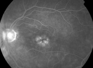
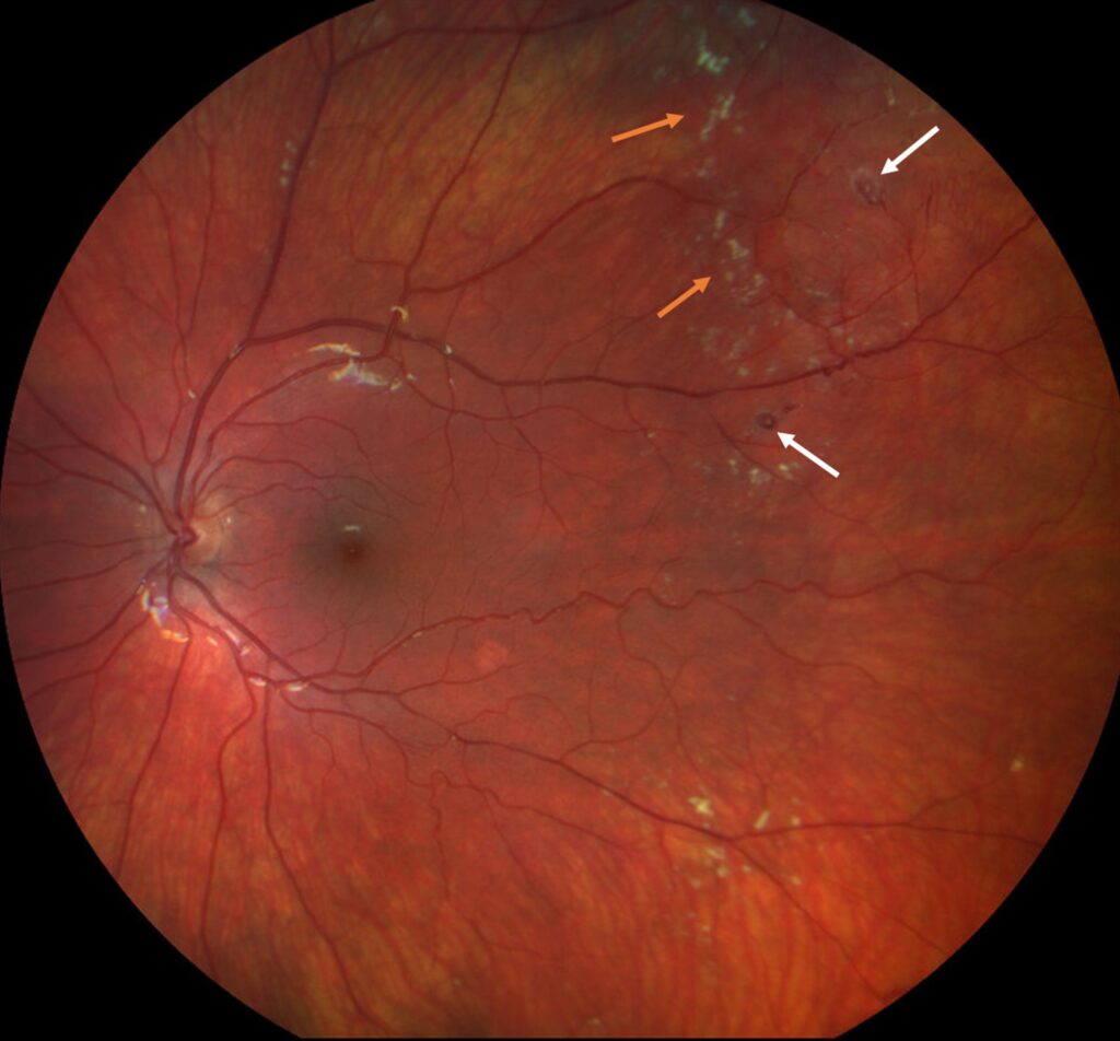
Fluorescein angiography highlights telangiectasia, aneurysmal changes, capillary dropout and leakage.
Meanwhile, OCT documents foveal exudate, subretinal fluid and fibrosis.
Ultrasound helps when the retina cannot be viewed.
Anti-VEGF Therapy in Coats Disease Stages
Intravitreal anti-VEGF agents can reduce exudation, particularly as an adjunct to ablation.
However, they do not replace definitive treatment of the abnormal vessels.
A small bevacizumab series also raised concern about vitreoretinal fibrosis and traction.
Therefore, clinicians should discuss this risk and monitor treated eyes carefully.
Visual Outlook by Coats Disease Stages
Higher stages generally carry worse vision and a greater risk of enucleation.
In the original Shields series, final acuity of 20/200 or worse rose steadily with stage.
No stage 1 eye reached that level, while all stage 4 and 5 eyes did.
Excluding Retinoblastoma Before Staging
Any child with leukocoria or exudative detachment needs retinoblastoma excluded.
Telangiectasia and yellow subretinal exudate favor Coats disease, whereas intraocular calcification suggests retinoblastoma.
Therefore, imaging should precede any drainage procedure when the diagnosis remains uncertain.
Another mimic is Coats-like retinal telangiectasia, which can accompany other conditions.
Other Mimics of Exudative Retinal Disease
First, familial exudative vitreoretinopathy is usually bilateral and shows peripheral avascular retina.
Also, a retinal capillary hemangioblastoma has a feeder vessel and may signal von Hippel-Lindau disease.
In addition, a Toxocara granuloma or persistent fetal vasculature can resemble advanced Coats disease.
Therefore, examine the fellow eye carefully before assigning a stage.
Examination Pearls for Coats Disease Stages
Because lesions often lie in the inferotemporal periphery, scleral depression is essential.
Also, wide-field photography documents the baseline extent for later comparison.
Then, check the fellow eye, since bilateral involvement is uncommon but possible.
As a result, staging stays consistent across visits.
Counseling Families About Coats Disease Stages
Families should understand that treatment aims to stop leakage and preserve the eye.
Vision may remain poor even after exudation clears, particularly with foveal scarring.
Finally, explain that repeated treatment sessions are common and that follow-up continues long term.
Follow-Up Across Coats Disease Stages
Early visits assess treatment response, residual telangiectasia and recurrent exudation.
Next, the interval can lengthen as leakage resolves.
Recurrence can occur after apparent control, so periodic dilated examinations remain necessary.
Pitfalls to Avoid
- Do not stage without examining the far peripheral retina
- Never drain subretinal fluid before excluding retinoblastoma
- Do not rely on anti-VEGF therapy alone
- Do not promise visual recovery after exudate resolves


Document what you see
Two smartphone imaging tools built for everyday clinic use — one for the slit lamp, one for the fundus.
From Choroida — the team behind this siteReferences
- Shields JA, Shields CL, Honavar SG, Demirci H, Cater J. Classification and management of Coats disease: the 2000 Proctor Lecture. Am J Ophthalmol. 2001;131:572-583.
- Shields JA, Shields CL, Honavar SG, Demirci H. Clinical variations and complications of Coats disease in 150 cases: the 2000 Sanford Gifford Memorial Lecture. Am J Ophthalmol. 2001;131:561-571.
- Sigler EJ, Randolph JC, Calzada JI, Charles S. Current management of Coats disease. Surv Ophthalmol. 2014;59:30-46.
- Ramasubramanian A, Shields CL. Bevacizumab for Coats’ disease with exudative retinal detachment and risk of vitreoretinal traction. Br J Ophthalmol. 2012;96:356-359.
- Shields CL, Udyaver S, Dalvin LA, et al. Visual acuity outcomes in Coats disease by classification stage in 160 patients. Br J Ophthalmol. 2020;104:422-431.

Writes and reviews the clinical articles on Educate. Every post is checked against peer-reviewed sources before it goes live.