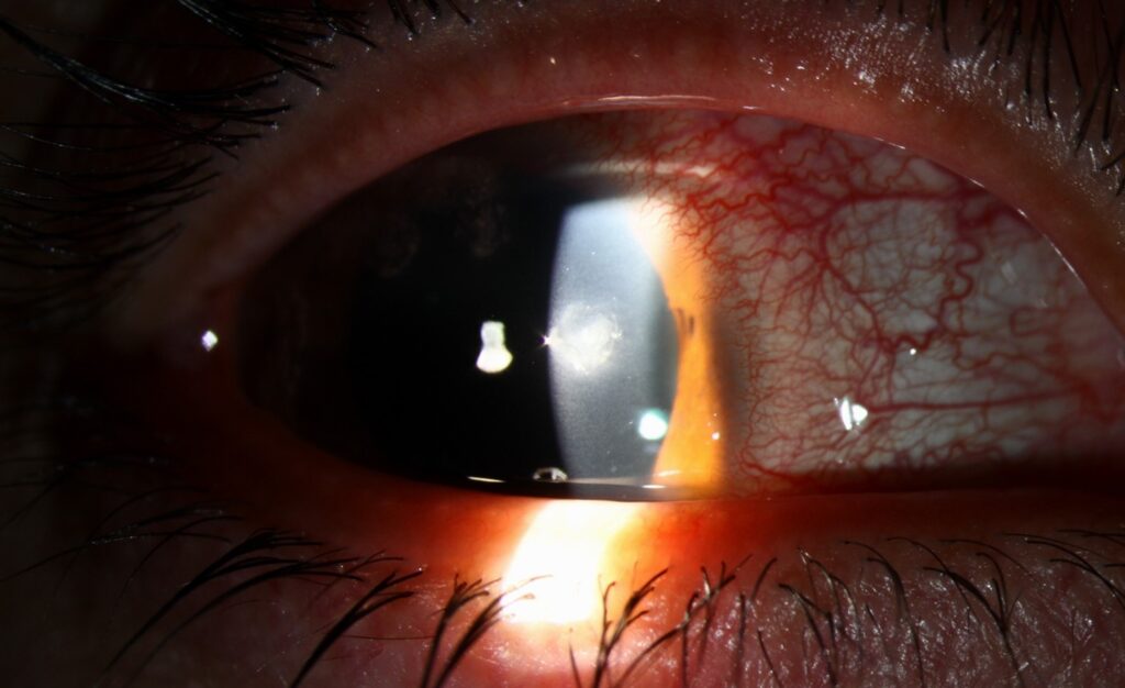
Ciliary flush is a violaceous ring of injection that hugs the limbus and fades toward the fornices.
Clinicians also call it circumcorneal injection.
The sign points to inflammation of the cornea, iris or ciliary body.
Therefore, it should always prompt a careful slit-lamp examination.
What Ciliary Flush Means
Ciliary flush reflects dilation of the deep anterior ciliary vessels near the limbus.
These vessels supply the iris, ciliary body and peripheral cornea.
As a result, inflammation in these tissues congests the limbal plexus.
The redness therefore sits deep to the conjunctiva.
Anatomy Behind the Sign
The anterior ciliary arteries travel with the rectus muscles and pierce the sclera near the limbus.
When the anterior uvea or cornea becomes inflamed, this deep network engorges.
Then the flush appears as a narrow band around the cornea.
All-fit Slit-Lamp Adapter
Record and share exactly what you see at the slit lamp. One adapter fits any slit lamp or surgical microscope — and any smartphone.
From Choroida — the team behind this siteHow Ciliary Flush Looks
The color is dusky red or violet.
Its vessels look straight, fine and radially arranged.
In addition, the redness is most intense at the limbus.
It fades toward the fornices, where the conjunctiva may look almost normal.
Conjunctival Injection vs Ciliary Flush
Conjunctival injection shows the opposite distribution.
It is bright red, most marked in the fornices and lighter near the limbus.
Its vessels are tortuous and move with the conjunctiva when you push the lower lid.
Also, they blanch with gentle pressure or a topical vasoconstrictor.
Episcleral Injection Compared
Episcleral vessels lie between the conjunctiva and the sclera.
Episcleritis usually causes sectoral, salmon-pink redness with mild discomfort.
However, these vessels often blanch after phenylephrine 10%.
In contrast, the deep scleral plexus in scleritis does not blanch and looks bluish.
Slit-Lamp Tests for Ciliary Flush
First, inspect the eye in daylight or diffuse white light, because color shows best this way.
Then instill topical phenylephrine and look again after several minutes.
Deep ciliary vessels stay red while superficial conjunctival vessels fade.
For technique, review diffuse and direct illumination.
Symptoms That Accompany the Flush
Patients usually report pain, photophobia and blurred vision.
By comparison, conjunctivitis causes grittiness and discharge with normal vision.
Therefore, pain plus reduced acuity raises the level of concern immediately.
Acute Anterior Uveitis and Ciliary Flush
Iritis is the classic cause of ciliary flush.
The pupil is often small and sluggish because of sphincter spasm.
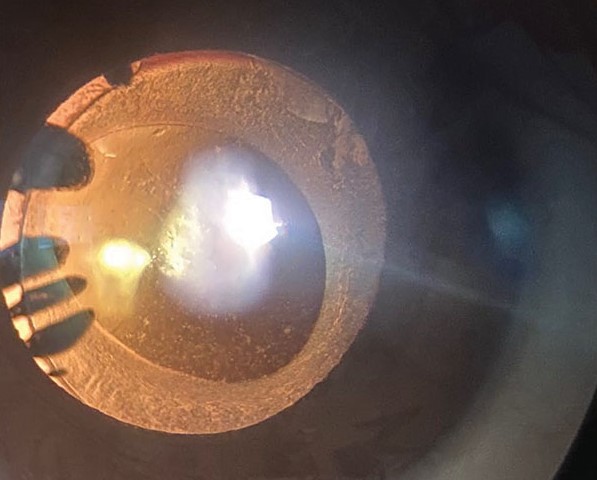
Look for anterior chamber cells and flare, keratic precipitates and posterior synechiae.
Also, consensual photophobia supports the diagnosis, as Au and Henkind described.
Keratitis and Corneal Injury
Microbial keratitis, herpes simplex keratitis and corneal abrasion can all produce the sign.
Fluorescein staining reveals the epithelial defect or dendrite.
An infiltrate with an overlying defect suggests infection until proved otherwise.
In addition, contact lens wear raises suspicion of bacterial or Acanthamoeba disease.
Acute Angle Closure
Angle closure causes a flush with severe pain, haloes, headache and nausea.
The cornea looks hazy from epithelial edema.
Meanwhile, the pupil is mid-dilated and poorly reactive, and the chamber is shallow.
Measure the pressure at once (see acute angle-closure glaucoma).
Other Causes of Ciliary Flush
- Scleritis, especially the diffuse anterior form
- Endophthalmitis after surgery or injection
- Corneal foreign body or chemical injury
- Lens-induced uveitis
- Traumatic iritis after blunt injury
Triage of Ciliary Flush
Treat ciliary flush as an urgent sign.
First, record visual acuity and check the pupils.
Next, measure intraocular pressure and stain the cornea with fluorescein.
Then examine the anterior chamber for cells, hypopyon and depth.
Finally, refer the same day when the cause is unclear or vision is reduced.
For the broader approach, see red eye triage.
Management Depends on Cause
The flush itself needs no treatment.
Instead, treatment targets the underlying disease.
For example, iritis needs topical corticosteroids and cycloplegia once infection is excluded.
Keratitis needs cultures and antimicrobials, while angle closure needs pressure lowering and iridotomy.
Vasoconstrictor drops only mask the sign and delay diagnosis.
Following Ciliary Flush Over Time
The flush usually fades as inflammation settles.
However, grade the chamber reaction and corneal findings, because these guide steroid tapering.
A persistent flush suggests poor control or a wrong diagnosis, such as scleritis or masked infection.
Counseling the Patient
Explain that deep redness around the cornea signals inflammation inside the eye.
Also, stress that it differs from a simple pink eye.
Ask patients to return immediately if pain or blurring worsens.
Pitfalls to Avoid
- Do not label a painful red eye with reduced vision as conjunctivitis
- Never start topical steroids before excluding herpetic or microbial keratitis
- Do not skip pressure measurement when the cornea looks hazy
- Avoid judging color under the yellow slit-lamp beam alone
For the latest studies, see the PubMed results for ciliary flush.


Document what you see
Two smartphone imaging tools built for everyday clinic use — one for the slit lamp, one for the fundus.
From Choroida — the team behind this siteReferences
- Leibowitz HM. The red eye. N Engl J Med. 2000;343:345-351.
- Cronau H, Kankanala RR, Mauger T. Diagnosis and management of red eye in primary care. Am Fam Physician. 2010;81:137-144.
- Watson PG, Hayreh SS. Scleritis and episcleritis. Br J Ophthalmol. 1976;60:163-191.
- Jabs DA, Nussenblatt RB, Rosenbaum JT. Standardization of uveitis nomenclature for reporting clinical data. Am J Ophthalmol. 2005;140:509-516.
- Au YK, Henkind P. Pain elicited by consensual pupillary reflex: a diagnostic test for acute iritis. Lancet. 1981;2:1254-1255.
- Bowling B. Kanski’s Clinical Ophthalmology: A Systematic Approach. 8th ed. Elsevier; 2016.

Writes and reviews the clinical articles on Educate. Every post is checked against peer-reviewed sources before it goes live.