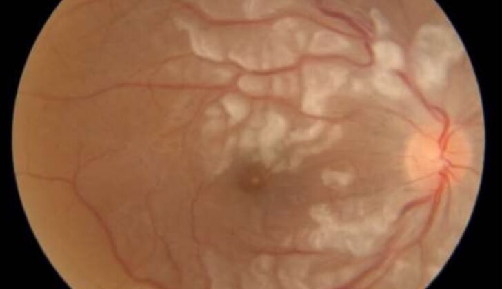
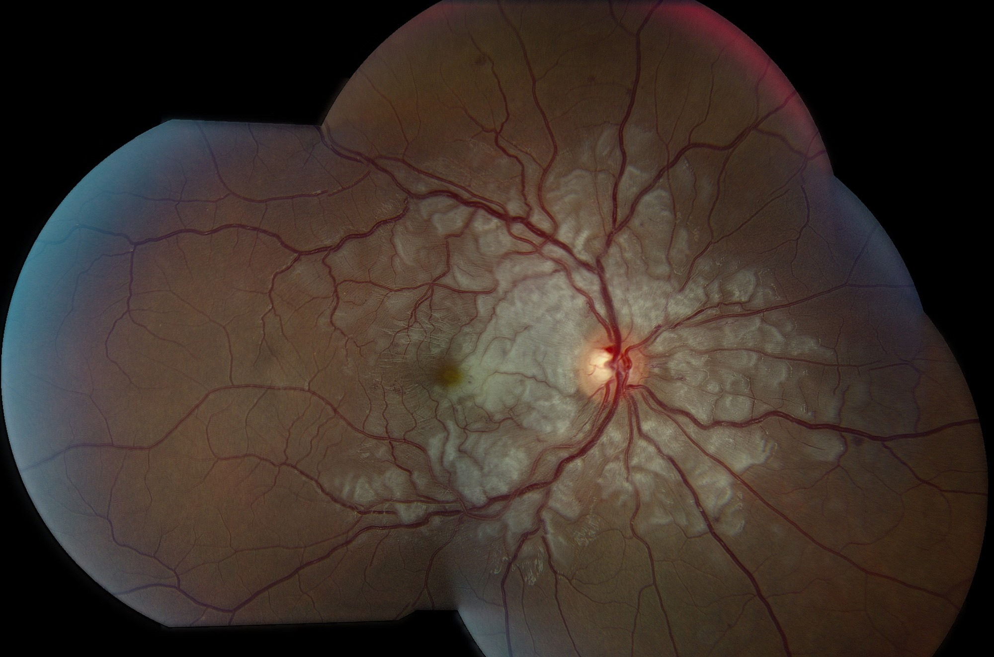
Purtscher flecken are polygonal patches of inner retinal whitening that lie between retinal arterioles and venules.
A narrow band of normal-looking retina separates each patch from the adjacent vessel.
Because this strip is so characteriztic, many authors regard it as the most specific fundus sign of Purtscher retinopathy.
However, clinicians often mistake the sign for cotton wool spots, which delays the search for a systemic cause.
What Purtscher Flecken Look Like
The patches appear gray-white with sharp, angular borders, mostly in the posterior pole.
Their shape follows the spaces between vessels, so they form a mosaic of irregular polygons.
In addition, a thin clear strip runs along both sides of the arterioles and venules.
The Perivascular Clear Zone
This clear strip is the defining feature of the sign.
Retina beside a patent vessel probably receives oxygen by diffusion from it, so it stays transparent.
However, this explanation is still a hypothesis.
Fundus Explorer Pro
Photograph the retinal findings described here with the phone already in your pocket — 22 D optics and built-in illumination in one handheld unit.
From Choroida — the team behind this siteWhy Purtscher Flecken Form
Most authors attribute the whitening to occlusion of precapillary arterioles.
For example, proposed emboli include complement-activated leukocyte aggregates, fat, fibrin and air.
As a result, the inner retina develops ischemic intracellular edema, which looks white.
Purtscher Flecken vs Cotton Wool Spots
Cotton wool spots are nerve fiber layer infarcts with fluffy, feathery edges (see cotton wool spots).
They also tend to sit over vessels and partly hide them.
Purtscher flecken, by contrast, have crisp polygonal margins and spare the retina beside the vessels.
Both signs can coexist, so inspect the border of every white lesion.
Associated Retinal Findings
Purtscher flecken rarely appear alone.
Cotton wool spots, flame hemorrhages and mild disc edema usually accompany them.
Some eyes also show macular whitening with a pseudo cherry red spot.
Exam Tips for Purtscher Flecken
Examine the posterior pole through a dilated pupil with a high-magnification lens.
Next, trace each patch to its nearest vessel and look for the clear strip.
Also, photograph the fundus early, because the flecken fade within weeks.
Causes of Purtscher Flecken
Otmar Purtscher described the picture in 1910 after severe head trauma.
Later reports, however, linked identical findings to many nontraumatic conditions:
- Head or chest compression injury
- Long bone fractures with fat embolism
- Acute pancreatitis
- Renal failure
- Systemic lupus erythematosus and other connective tissue diseases
- Preeclampsia, HELLP syndrome and childbirth
- Thrombotic thrombocytopenic purpura and hemolytic uremic syndrome
Diagnostic Criteria
Miguel and colleagues proposed criteria in a 2013 systematic review.
Their criteria require three of five features, starting with flecken, few to moderate flame hemorrhages, and posterior pole cotton wool spots.
Finally, they add a plausible cause and compatible investigations (see Purtscher retinopathy).
OCT in Purtscher Flecken
Acute OCT shows thickened, hyperreflective inner retinal layers within the whitened areas.
The spared perivascular retina keeps its normal layered profile.
Later scans, however, often show inner retinal thinning, which reflects the ischemic injury.
Angiography Findings
Fluorescein angiography may show arteriolar nonperfusion and capillary dropout in affected zones.
Leakage and late staining of damaged vessels can also appear.
OCT angiography maps capillary nonperfusion without dye, although findings vary with timing.
Differential Diagnosis of Purtscher Flecken
Commotio retinae after blunt trauma whitens the outer retina at the impact site.
By contrast, artery occlusion whitens a whole vascular territory (see branch retinal artery occlusion).
Finally, hypertensive and diabetic cotton wool spots lack both the polygonal shape and the clear zone.
Systemic Workup
Ask about trauma, abdominal pain, pregnancy, alcohol use, and kidney or autoimmune disease.
Then, work with the medical team on lipase, renal function, and a blood count with smear.
Because thrombotic microangiopathy can be life-threatening, treat an abnormal smear as urgent.
Management of Purtscher Flecken
No therapy acts directly on the flecken, so treatment targets the systemic disease.
Some clinicians give high-dose corticosteroids, based on case reports and small series.
However, no controlled trial supports this approach, and reported outcomes after observation look broadly similar.
Prognosis and Follow-Up
The whitening usually fades over weeks to a few months.
Visual outcome, however, depends on macular and optic nerve damage and ranges from full recovery to permanent loss.
Late signs include disc pallor, pigment mottling and inner retinal thinning on OCT.
Counseling Patients
Explain that the white patches reflect small blocked retinal vessels.
They fade with time, although lost vision may not fully return.
Also, the eye findings point to a systemic problem that needs medical care.
Pitfalls to Avoid
- Labeling every white patch a cotton wool spot without checking borders
- Missing the clear zone at low magnification
- Delaying photography until the flecken fade
- Overlooking pancreatitis in a patient without trauma
- Promising recovery before the macula and disc stabilize


Document what you see
Two smartphone imaging tools built for everyday clinic use — one for the slit lamp, one for the fundus.
From Choroida — the team behind this siteReferences
- Purtscher O. Noch unbekannte Befunde nach Schädeltrauma. Ber Dtsch Ophthalmol Ges. 1910;36:294-301.
- Agrawal A, McKibbin MA. Purtscher’s and Purtscher-like retinopathies: a review. Surv Ophthalmol. 2006;51:129-136.
- Agrawal A, McKibbin M. Purtscher’s retinopathy: epidemiology, clinical features and outcome. Br J Ophthalmol. 2007;91:1456-1459.
- Miguel AIM, Henriques F, Azevedo LFR, Loureiro AJR, Maberley DAL. Systematic review of Purtscher’s and Purtscher-like retinopathies. Eye. 2013;27:1-13.
- American Academy of Ophthalmology. Basic and Clinical Science Course, Section 12: Retina and Vitreous. San Francisco: AAO; 2023.

Writes and reviews the clinical articles on Educate. Every post is checked against peer-reviewed sources before it goes live.