
A Weiss ring is a ring-shaped floater that patients describe as a circle, a “ring,” a “hair,” or a “spider web.” It is the margin of vitreous attachment around the optic disc, which has peeled away when the vitreous gel detached from the retina.
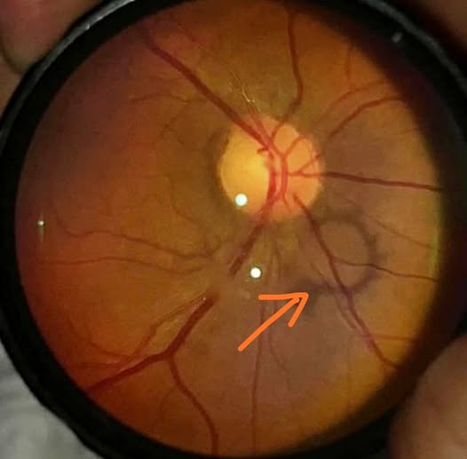

If you can see one on slit-lamp examination, the vitreous has detached completely from the disc.
Why it forms
At the optic disc, the vitreous is attached to a ring of glial tissue around the disc margin.
When the vitreous separates from the retina, it pulls this glial tissue away.
The tissue floats in the posterior vitreous cavity as a round or oval ring, sometimes broken, often accompanied by wisps of gel.
The ring is named after Martin Weiss, who described it in the 1930s, and it is almost always seen with a posterior vitreous detachment (see posterior vitreous detachment).
Symptoms
- A sudden new floater, often described as a ring, a circle, or a cobweb in the field of view
- Flashes of light in the temporal field, caused by traction at the vitreoretinal adhesion
- Sometimes symptoms are minimal, and the ring is found incidentally
- The floater often moves with eye movements and tends to settle then drift, being most obvious against a bright background
Fundus Explorer Pro
Photograph the retinal findings described here with the phone already in your pocket — 22 D optics and built-in illumination in one handheld unit.
From Choroida — the team behind this siteWhy it matters
The ring itself is harmless.
The significance is that it marks a recent posterior vitreous detachment, and in a minority of eyes the detachment causes a retinal tear.
The risk is highest in the first six weeks and in patients with symptoms such as flashes, a shower of new floaters, or a visual field shadow (see vitreous floaters and lattice degeneration).
Examination
- Dilated examination with scleral depression of the entire periphery
- Slit-lamp biomicroscopy of the vitreous, looking for pigment cells in the anterior vitreous (Shafer sign), which suggests a tear
- Vitreous hemorrhage or pigment cells call for a detailed search for breaks
- Document the retina carefully and explain warning symptoms before sending the patient home
Advice for patients
- Floaters usually become less noticeable over weeks to months as the brain adapts
- Return urgently for new flashes, a sudden increase in floaters, or a shadow or curtain in the vision
- A second examination after about four to six weeks is reasonable when the first exam was normal but symptoms were acute
What the examiner is looking for
The risk of a retinal tear after acute symptomatic PVD is around 10 to 15 percent in most series, and it rises sharply when there is vitreous hemorrhage or pigment in the vitreous.
Tears are usually found in the superior retina, where the vitreous exerts the most traction when the eye moves.
A careful peripheral examination, usually with scleral depression, is the reason for the visit, and the Weiss ring is only the reason the patient came in.
When the exam is normal, a patient should be told clearly that the risk is not zero.
About 2 to 5 percent of patients develop new breaks at a follow-up visit in the next weeks, and a second look is justified in patients with persistent flashes or a high-risk profile such as high myopia, previous retinal detachment in the fellow eye, or a family history.
Treatment
No treatment is needed for the ring.
Any retinal tear is treated with laser retinopexy or cryotherapy.
Vitrectomy for floaters is reserved for patients with severe, persistent symptoms who understand the risks, and YAG vitreolysis is used by some surgeons with variable results.
What patients actually describe
Many people say that they see "a fly", "a cobweb", or "a circle that follows my eye". A Weiss ring is often described as a floating ring or a rounded, semi-transparent shape, and it is most noticeable against a bright, plain background such as a wall or the sky. Some patients can even draw it. Asking the patient to sketch the shape and tell you the side on which it first appeared is a useful way to confirm the history.
When to repeat the examination
Repeat the dilated examination if the symptoms change, if new flashes appear, or if a veil or shadow develops. A second visit in a few weeks is sensible in a high-risk patient, since some breaks develop after the first examination. Explain that the floater may persist and that most people stop noticing it as the brain learns to ignore it.
Prognosis
Most patients do very well, and the floater becomes less intrusive with time.


Document what you see
Two smartphone imaging tools built for everyday clinic use — one for the slit lamp, one for the fundus.
From Choroida — the team behind this siteReferences
- Hollands H, Johnson D, Brox AC, Almukhtar T, Sharma S, Hollands S. Acute-onset floaters and flashes: is this patient at risk for retinal detachment? JAMA. 2009;302:2243-2249.
- Johnson MW. Posterior vitreous detachment: evolution and complications of its early stages. Am J Ophthalmol. 2010;149:371-382.
- Coffee RE, Westfall AC, Davis GH, Mieler WF, Holz ER. Symptomatic posterior vitreous detachment and the incidence of delayed retinal breaks. Am J Ophthalmol. 2007;144:409-413.
- Foos RY, Wheeler NC. Vitreoretinal juncture; synchysis senilis and posterior vitreous detachment. Ophthalmology. 1982;89:1502-1512.
