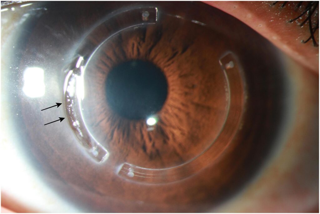
Intracorneal ring segments (ICRS) are small arc-shaped implants made of polymethyl methacrylate that are placed in a tunnel within the corneal stroma to flatten and regularize the cornea.
Clinicians use them in keratoconus and in ectasia after refractive surgery, mostly for patients who can no longer wear contact lenses comfortably.
They improve the shape of the cornea, but they do not stop progression on their own.
What Are Intracorneal Ring Segments?
The segments are placed in the mid-peripheral stroma, and they add material to the periphery of the cornea.
Also, by the principle of arc shortening, this flattens the central cornea, and the amount of flattening depends on the thickness and the arc length of the segments.
Several designs exist, including Intacs, Ferrara rings, and Keraring, with different cross-sections and arc lengths.
Asymmetric cones, with the steep area off center, are treated with segments of different thickness above and below the apex, according to a nomogram.
Clinicians create the tunnel with a mechanical dissector or with a femtosecond laser, which gives better depth control.
Indications of Intracorneal Ring Segments
- Keratoconus with contact lens intolerance and spectacle blur
- Clear central cornea without scarring in the visual axis
- Adequate corneal thickness at the planned site of the tunnel, usually not less than about 450 micrometers
- Moderate keratometry, since extremely steep corneas respond less predictably
- Post-LASIK or post-PRK ectasia (see post-LASIK ectasia)
- Pellucid marginal degeneration in selected cases (see pellucid marginal degeneration)
Patients with central scars, a very thin cornea, or severe disease may be better served by other treatments such as keratoplasty.
All-fit Slit-Lamp Adapter
Record and share exactly what you see at the slit lamp. One adapter fits any slit lamp or surgical microscope — and any smartphone.
From Choroida — the team behind this siteIntracorneal Ring Segments: Combination With Cross-Linking
Because segments do not strengthen the cornea, the underlying disease can continue to progress.
Many surgeons therefore combine the implants with corneal cross-linking, either at the same time or sequentially (see corneal collagen cross-linking).
Studies suggest that the combination gives more stable flattening and a lower rate of progression than segments alone, although the best sequence and timing remain debated.
Expected Results
In published series, ICRS reduce the steepest keratometry by several diopters on average.
They improve uncorrected and best-corrected acuity in many patients.
The effect varies between eyes, and some patients gain a few lines while others have little change.
Contact lens tolerance often improves, and a patient who could not wear lenses may be able to do so again.
The improvement may continue to evolve for months as the cornea remodels.
Complications of Intracorneal Ring Segments
- Segment migration or extrusion, especially if shallow or near the incision
- Infectious keratitis around the tunnel, a rare but serious event
- Corneal thinning, melting, and stromal deposits near the segments
- Glare, halos, and induced astigmatism
- Neovascularization and persistent discomfort
- Poor centration and undercorrection or overcorrection
Segments can be removed or exchanged if there is a complication, and the cornea usually returns toward its previous shape.
Counselling
Explain that the aim is to improve vision and contact lens tolerance and to delay or avoid a corneal transplant, not to restore normal vision.
Make clear that a cross-linking procedure may also be needed to stabilize the disease.
Discuss the possible need for glasses or lenses after surgery, and the small risks of infection and migration.
Planning the Procedure
The surgeon uses topography and tomography to locate the cone, to measure the corneal thickness along the planned tunnel, and to choose segment thickness and arc length from a nomogram.
Patients with a cone in the inferior cornea and a symmetric bow tie may receive one or two segments, while those with a central or asymmetric cone may receive different configurations.
Consider the patient’s contact lens history, the stability of keratometry, and the status of the fellow eye.
The Procedure Itself
Clinicians perform the operation under topical anesthesia, with a marked center, a radial incision at the planned meridian, and creation of a tunnel at a depth of about two thirds of the thickness.
The segments are inserted and positioned, and the incision is closed with a suture in some techniques.
Postoperative care includes topical antibiotics and steroids for a short period, with review in a day, a week, and a month.
Long-Term Follow-Up
Examine the segments periodically for depth, deposits, and any thinning of the overlying stroma.
Obtain topography at intervals to document the effect and to see whether the disease is still progressing.
If progression continues despite segments, plan cross-linking or other treatment without delay.
Informed consent should cover the chance that the implants fail to improve vision, the possibility of removal, and the need for continued monitoring of the disease.
Give patients the contact details of the clinic for any redness or pain after the operation, since infection around the segments needs immediate treatment.
For the latest studies, see the PubMed results for intracorneal ring segments.


Document what you see
Two smartphone imaging tools built for everyday clinic use — one for the slit lamp, one for the fundus.
From Choroida — the team behind this siteReferences
- Piñero DP, Alio JL. Intracorneal ring segments in ectatic corneal disease: a review. Clin Exp Ophthalmol. 2010;38:154-167.
- Colin J, Cochener B, Savary G, Malet F. Correcting keratoconus with intracorneal rings. J Cataract Refract Surg. 2000;26:1117-1122.
- Gomes JAP, Tan D, Rapuano CJ, et al. Global consensus on keratoconus and ectatic diseases. Cornea. 2015;34:359-369.
- American Academy of Ophthalmology. Basic and Clinical Science Course, Section 8: External Disease and Cornea. AAO; current edition.

Writes and reviews the clinical articles on Educate. Every post is checked against peer-reviewed sources before it goes live.