Posterior uveitis is intraocular inflammation centered in the retina, the choroid or both.
The SUN Working Group defines it by anatomic site, so vitritis alone does not qualify.
Patients usually report floaters, blurred vision or scotomas, while the anterior segment may look quiet.
Therefore, a dilated fundus examination decides the diagnosis.
What Counts as Posterior Uveitis
Inflammation qualifies when the primary site is the retina or choroid.
Retinitis, choroiditis, retinochoroiditis and retinal vasculitis all fall within this group.
In contrast, inflammation centered in the vitreous and pars plana is intermediate uveitis.
When all segments are involved equally, the term panuveitis applies.
Symptoms That Bring Patients In
Floaters and painless blurring are the most common complaints.
Photopsia, paracentral scotomas and distorted vision suggest outer retinal or macular involvement.
However, peripheral lesions may cause no symptoms at all.
Pain and redness are less typical, so their presence raises suspicion of anterior spillover or scleritis.
Fundus Explorer Pro
Photograph the retinal findings described here with the phone already in your pocket — 22 D optics and built-in illumination in one handheld unit.
From Choroida — the team behind this siteExam Signs in Posterior Uveitis
- Vitreous cells and haze, graded with standardized photographs
- Creamy or yellow choroidal lesions, focal or multifocal
- Fluffy white retinitis with indistinct borders
- Perivascular sheathing from retinal vasculitis
- Macular edema, disc swelling or serous retinal detachment
Infectious Versus Noninfectious Causes
The first clinical decision is whether an organism could be driving the inflammation.
Missing an infection is dangerous, because corticosteroids alone can worsen it.
Therefore, treat every new case as infectious until history, exam and tests suggest otherwise.
Infectious Causes of Posterior Uveitis
- Toxoplasmosis, the most common cause of retinochoroiditis in many regions
- Syphilis, which can mimic almost any pattern
- Tuberculosis, often as serpiginous-like or multifocal choroiditis
- Herpes viruses causing acute retinal necrosis
- Cytomegalovirus retinitis in immunosuppressed patients
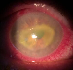
- Endogenous bacterial or fungal endophthalmitis
Syphilis also produces a distinct placoid lesion (see syphilitic placoid chorioretinitis).
Noninfectious Causes
Sarcoidosis and Behçet disease are major systemic causes.
Also, Vogt-Koyanagi-Harada disease and sympathetic ophthalmia present with bilateral granulomatous choroiditis.
In addition, the white dot syndromes form a large eye-limited group.
Focal Patterns in Posterior Uveitis
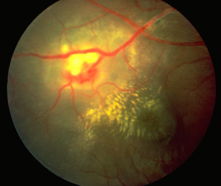
A single active lesion points toward a short list.
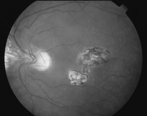
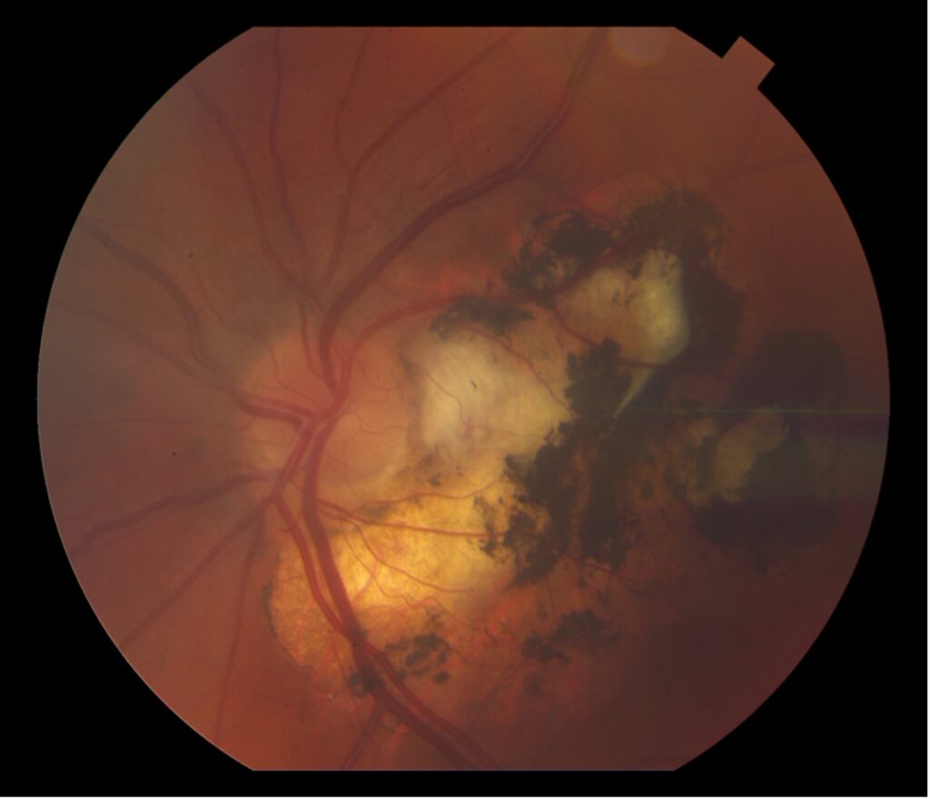
For example, focal retinochoroiditis next to a pigmented scar strongly suggests toxoplasmosis.
A solitary choroidal granuloma raises tuberculosis or sarcoidosis.
Then, an isolated subretinal abscess in an unwell patient suggests endogenous infection.
Multifocal Patterns
Multiple lesions widen the differential.
Small punched-out lesions in a young myopic woman suggest multifocal choroiditis (see multifocal choroiditis and panuveitis).
Cream-colored ovoid spots around the disc fit birdshot chorioretinopathy.
Retinal Vasculitis
Sheathing or leakage of retinal vessels needs its own differential.
Occlusive venous vasculitis suggests Behçet disease, while segmental periphlebitis may point to sarcoidosis or tuberculosis.
Arterial involvement raises concern for viral retinitis or systemic vasculitis.
Imaging in Posterior Uveitis
Angiography with fluorescein shows vascular leakage, macular edema and active lesion borders.
Indocyanine green angiography reveals choroidal lesions hidden on examination.
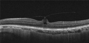
Meanwhile, OCT measures macular thickness and shows outer retinal damage.
Stepwise Workup for Posterior Uveitis
- Take a detailed history: travel, sexual risk, TB exposure, pets, immune status and systemic symptoms.
- Examine both eyes and classify the anatomic site, laterality and pattern.
- Order syphilis serology and an interferon-gamma release assay in most patients.
- Add a chest image, ACE or HLA-A29 only when the pattern supports them.
- Sample aqueous or vitreous for PCR when infection is suspected and the picture is unclear.
Masquerade Syndromes
Older patients with vitritis that resists steroids need special caution.
Vitreoretinal lymphoma can imitate inflammation for months (see masquerade syndromes).
Therefore, consider diagnostic vitrectomy when the course looks atypical.
Treatment of Infectious Posterior Uveitis
Antimicrobial therapy targets the organism directly.
For example, acute retinal necrosis needs prompt antiviral therapy, and ocular syphilis is treated as neurosyphilis.
Oral corticosteroids may be added later to limit inflammatory damage, but only under antimicrobial cover.
However, treatment cannot reverse retinal necrosis or scars that already exist.
Treatment of Noninfectious Posterior Uveitis
Corticosteroids control acute inflammation, given orally, periocularly or intravitreally.
Then, steroid-sparing immunosuppression is usually needed for chronic or bilateral disease.
The VISUAL I trial showed that adalimumab reduced the risk of treatment failure in active noninfectious uveitis.
Follow-Up and Complications
Review activity with vitreous haze grade, OCT thickness and angiographic leakage.
In addition, watch for macular edema, choroidal neovascularization, glaucoma and cataract.
Pitfalls to Avoid
- Do not start high-dose steroids before excluding syphilis and tuberculosis
- Never assume a quiet anterior chamber means a quiet fundus
- Do not label steroid-resistant vitritis in older patients as idiopathic
- Avoid ordering every test for every patient, because false positives mislead


Document what you see
Two smartphone imaging tools built for everyday clinic use — one for the slit lamp, one for the fundus.
From Choroida — the team behind this siteReferences
- Jabs DA, Nussenblatt RB, Rosenbaum JT; SUN Working Group. Standardization of uveitis nomenclature for reporting clinical data. Am J Ophthalmol. 2005;140:509-516.
- Standardization of Uveitis Nomenclature (SUN) Working Group. Development of classification criteria for the uveitides. Am J Ophthalmol. 2021;228:96-105.
- Jabs DA, Rosenbaum JT, Foster CS, et al. Guidelines for the use of immunosuppressive drugs in patients with ocular inflammatory disorders. Am J Ophthalmol. 2000;130:492-513.
- Jaffe GJ, Dick AD, Brézin AP, et al. Adalimumab in patients with active noninfectious uveitis. N Engl J Med. 2016;375:932-943.
- American Academy of Ophthalmology. Basic and Clinical Science Course, Section 9: Uveitis and Ocular Inflammation. San Francisco: AAO.

Writes and reviews the clinical articles on Educate. Every post is checked against peer-reviewed sources before it goes live.