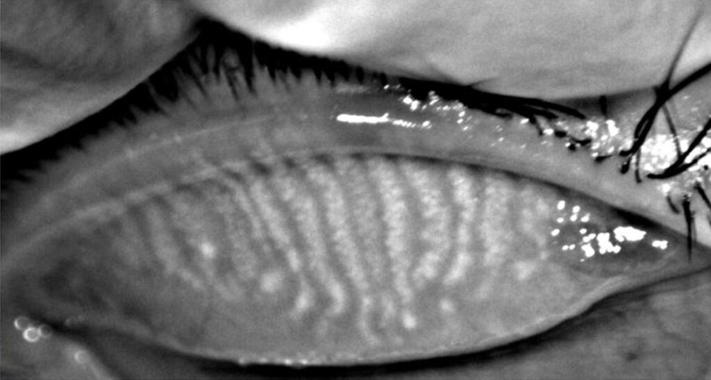
Meibography images the meibomian glands directly, usually with infrared light through the everted eyelid.
It shows gland structure that slit lamp examination cannot see.
As a result, it has become a routine part of many dry eye clinics.
What Meibography Shows
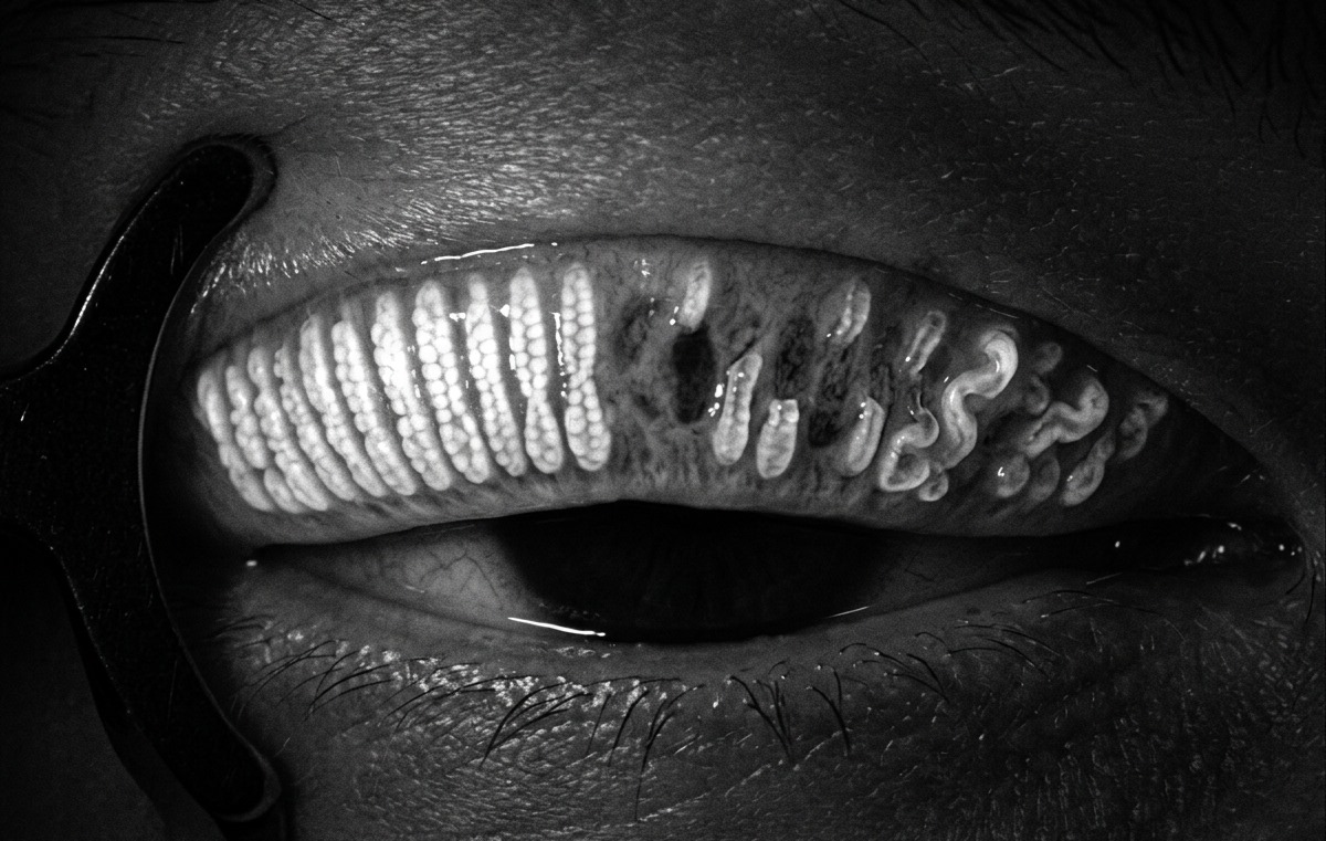
Each gland appears as a bright, elongated band on a darker tarsal background.
Meibography therefore reveals dropout, shortening, tortuosity and dilation of individual glands.
However, it shows structure only and says little about secretion quality.
How Infrared Meibography Works
Near-infrared light penetrates the tarsal conjunctiva and reflects from lipid-filled acini.
Older transillumination systems placed a light source against the skin side of the lid.
In contrast, noncontact systems image the everted lid directly, which improves comfort and speed.
All-fit Slit-Lamp Adapter
Record and share exactly what you see at the slit lamp. One adapter fits any slit lamp or surgical microscope — and any smartphone.
From Choroida — the team behind this siteTechnique at the Slit Lamp
First, explain the procedure, because lid eversion can feel unpleasant.
Next, evert the upper lid fully and keep the tarsal plate flat.
Then pull the lower lid down while the patient looks up.
Finally, capture the whole tarsus of both lids in each eye.
Common Errors in Meibography
Incomplete eversion hides the proximal glands and mimics shortening.
Also, reflection from the tear film can wash out part of the image.
Therefore, repeat any image that does not show the full tarsus in focus.
Normal Findings and Aging
Arita and colleagues documented meibomian gland loss in normal subjects with noncontact infrared meibography.
Dropout increased with age in that population.
As a result, mild partial loss in an older adult does not prove disease on its own.
Grading Gland Dropout in Meibography
Grading converts an image into a number that you can track over time.
Most scales estimate the proportion of tarsal area with gland loss.
Subjective scales are fast, while computer analysis gives a continuous percentage.
The Arita Meiboscore
The meiboscore grades each lid from 0 to 3.
Grade 0 means no loss, while grade 1 means loss of less than one third of the gland area.
Grade 2 covers loss between one third and two thirds, and grade 3 covers more than two thirds.
Then add the two lids for a total score from 0 to 6.
The Pult Meiboscale
The Pult meiboscale uses five grades, from 0 to 4, in steps of roughly one quarter.
Pult and Riede-Pult compared this subjective grading with computerized analysis of the same images.
However, scores from different scales are not interchangeable, so use one scale for each patient.
Gland Morphology Beyond Dropout
Shortened glands that stop short of the lid margin suggest early atrophy.
Also, tortuous or dilated glands may reflect obstruction and raised intraductal pressure.
These changes can appear before obvious dropout.
Meibography in the Dry Eye Workup
TFOS DEWS II includes meibography among tests that help characterize the evaporative component of dry eye.
However, structure and function often disagree, so always combine imaging with gland expression.
A gland that looks intact may still secrete thick, toothpaste-like meibum.
For the wider clinical picture, see our review of dry eye disease.
Associations Seen in Meibography
Gland loss on imaging has been linked with several factors:
- Increasing age
- Contact lens wear
- Allergic and atopic eye disease
- Systemic isotretinoin
- Demodex blepharitis
- Long-term topical glaucoma therapy
Counseling With Meibography Images
Patients often understand their condition faster when they see their own glands.
For example, a picture of missing glands explains why drops alone give short relief.
The image can also improve adherence to warm compresses and lid hygiene.
However, avoid alarming language, because mild loss is common with age.
How Gland Loss Changes Treatment
Gland atrophy is generally regarded as irreversible, so treatment aims to protect the remaining glands.
Patients with good gland reserve often respond to heat, expression and lid hygiene (see meibomian gland dysfunction).
In contrast, extensive loss limits what gland-directed therapy can achieve.
These patients may need more lubrication, anti-inflammatory therapy and attention to any aqueous-deficient dry eye.
Procedures and Meibography
Thermal pulsation, intense pulsed light and intraductal probing all target obstructed glands.
Some small studies describe improved gland appearance after treatment.
However, evidence for true gland regeneration is limited, so set modest expectations.
Follow-Up Meibography
Repeat imaging with the same device and lid technique, then grade each lid separately.
Also, image before cataract or refractive surgery, because surgery can destabilize the ocular surface.
Finally, focal loss beside a lid mass needs careful assessment for a tumor.
Pitfalls to Avoid
- Do not diagnose MGD from an image without expression
- Never compare scores from different grading scales
- Do not mistake incomplete eversion for gland shortening
- Do not promise that lost glands will grow back


Document what you see
Two smartphone imaging tools built for everyday clinic use — one for the slit lamp, one for the fundus.
From Choroida — the team behind this siteReferences
- Arita R, Itoh K, Inoue K, Amano S. Noncontact infrared meibography to document age-related changes of the meibomian glands in a normal population. Ophthalmology. 2008;115:911-915.
- Pult H, Riede-Pult B. Comparison of subjective grading and objective assessment in meibography. Cont Lens Anterior Eye. 2013;36:22-27.
- Pult H, Nichols JJ. A review of meibography. Optom Vis Sci. 2012;89:E760-E769.
- Wolffsohn JS, Arita R, Chalmers R, et al. TFOS DEWS II diagnostic methodology report. Ocul Surf. 2017;15:539-574.
- Nichols KK, Foulks GN, Bron AJ, et al. The International Workshop on Meibomian Gland Dysfunction: executive summary. Invest Ophthalmol Vis Sci. 2011;52:1922-1929.

Writes and reviews the clinical articles on Educate. Every post is checked against peer-reviewed sources before it goes live.