Aspergillus keratitis is a filamentary fungal infection of the cornea caused by species such as Aspergillus flavus and Aspergillus fumigatus.
In many tropical series, it ranks second to Fusarium among mold ulcers.
However, Aspergillus leads the isolates in some regions.
Because the clinical picture overlaps with other molds, the laboratory usually settles the diagnosis.
What Is Aspergillus Keratitis
Aspergillus is a saprophytic mold found in soil, decaying vegetation and grain dust.
It enters the stroma after an epithelial break, usually from trauma.
Then hyphae spread along the stromal lamellae toward Descemet membrane.
As a result, untreated ulcers may perforate or invade the anterior chamber.
Risk Factors for Aspergillus Keratitis
Trauma with plant material is the classic history, especially in agricultural workers.
- Corneal injury with vegetable matter or soil
- Hot, humid climates and harvest seasons
- Prior topical corticosteroid use
- Contact lens wear, although bacteria dominate this group
- Diabetes, ocular surface disease or prior corneal surgery
All-fit Slit-Lamp Adapter
Record and share exactly what you see at the slit lamp. One adapter fits any slit lamp or surgical microscope — and any smartphone.
From Choroida — the team behind this siteSymptoms Patients Report
Patients report pain, redness, tearing, photophobia and blurred vision.
Onset is often slower than in bacterial keratitis.
For example, a farmer may present one to two weeks after a minor scratch.
Slit Lamp Signs of Aspergillus Keratitis
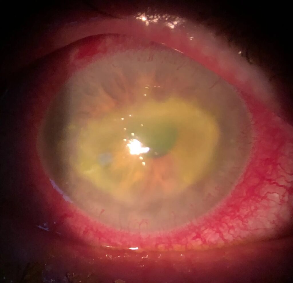
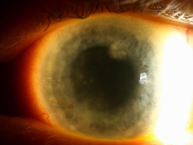
The infiltrate is typically gray-white with a dry, raised texture.
Its margins look feathery because hyphae extend into the surrounding stroma.
In addition, the examiner may see satellite lesions, an immune ring and an endothelial plaque.
A hypopyon is common and may appear thick and immobile.
Aspergillus Versus Fusarium at the Slit Lamp
No single sign reliably separates the two genera.
However, some series describe deeper and earlier stromal penetration with Fusarium.
Aspergillus ulcers may show a more elevated, dry slough, although this varies.
Therefore, treat morphology as a hint and confirm with smear and culture (see fungal vs bacterial keratitis clues).
Smear Findings
Scrape the leading edge and base of the ulcer with a blade or spatula.
Potassium hydroxide, calcofluor white and Gram stain all reveal fungal elements.
Aspergillus shows narrow, hyaline, septate hyphae with regular dichotomous branching at acute angles.
Fusarium hyphae look similar, so the smear confirms a mold while culture names the genus.
Technique details appear in corneal scraping and culture.
Culture in Aspergillus Keratitis
Sabouraud dextrose agar without cycloheximide is the standard medium.
Aspergillus often grows within two to three days.
Then colony color helps, with A. flavus yellow-green, A. fumigatus blue-green and A. niger black.
Colony microscopy shows conidiophores ending in a vesicle covered with phialides.
In contrast, Fusarium produces banana-shaped multiseptate macroconidia.
Differential Diagnosis
- Fusarium and other filamentary fungi
- Bacterial keratitis, especially partially treated ulcers
- Nocardia keratitis with a fungal-looking infiltrate
- Acanthamoeba keratitis
- Pythium keratitis in endemic regions
Natamycin in Aspergillus Keratitis
Natamycin 5% is the usual first-line topical agent for filamentary fungal keratitis.
MUTT I showed better outcomes with natamycin than topical voriconazole for filamentary ulcers overall (see MUTT results).
However, the benefit came mainly from Fusarium cases.
For non-Fusarium ulcers, including Aspergillus, the trial found no clear difference between drugs.
Also, natamycin penetrates deep stroma poorly.
Where Voriconazole Fits
Voriconazole 1% is a reasonable alternative or addition in Aspergillus keratitis.
In vitro studies often report good voriconazole activity against Aspergillus isolates.
It also penetrates the cornea better than natamycin.
Therefore, many clinicians add it for deep infiltrates or slow response.
Intrastromal injection is an option for deep disease, although evidence comes mostly from small series.
Oral Antifungals and Steroids
MUTT II found no overall benefit from adding oral voriconazole to topical therapy.
Systemic azoles also carry hepatic, visual and drug interaction risks.
As a result, reserve them for selected cases such as scleral or intraocular extension.
Avoid corticosteroids during active infection because they can promote fungal growth.
Monitoring Response
Review the patient daily or every few days in the early phase.
Record infiltrate size, epithelial defect, hypopyon height and thinning at each visit.
Because improvement is slow, judge the trend over several days.
Next, repeat the scraping if the ulcer worsens despite adequate therapy.
Surgical Triggers in Aspergillus Keratitis
Medical failure usually declares itself as progression in depth or width.
- Perforation or impending perforation
- Progressive infiltrate despite maximal medical therapy
- Extension toward the limbus or sclera
Therapeutic penetrating keratoplasty aims to remove all infected tissue with a clear margin.
However, graft reinfection and rejection remain real risks.
Small perforations may first receive tissue adhesive and a bandage contact lens.
Counseling in Aspergillus Keratitis
Explain that treatment lasts weeks and drops are frequent.
Also, warn that scarring may limit vision after the infection clears.
Patients should return at once if pain or redness increases.
Finally, advise protective eyewear during farm work.
Pitfalls to Avoid
- Starting steroids before ruling out fungus
- Swabbing the surface instead of scraping the ulcer base
- Stopping antifungals once the epithelium heals
- Assuming a negative smear excludes fungal infection


Document what you see
Two smartphone imaging tools built for everyday clinic use — one for the slit lamp, one for the fundus.
From Choroida — the team behind this siteReferences
- Prajna NV, Krishnan T, Mascarenhas J, et al. The Mycotic Ulcer Treatment Trial: a randomized trial comparing natamycin vs voriconazole. JAMA Ophthalmol. 2013;131:422-429.
- Prajna NV, Krishnan T, Rajaraman R, et al. Effect of oral voriconazole on fungal keratitis in the Mycotic Ulcer Treatment Trial II (MUTT II). JAMA Ophthalmol. 2016;134:1365-1372.
- Thomas PA, Kaliamurthy J. Mycotic keratitis: epidemiology, diagnosis and management. Clin Microbiol Infect. 2013;19:210-220.
- Thomas PA. Fungal infections of the cornea. Eye. 2003;17:852-862.
- Leck AK, Thomas PA, Hagan M, et al. Aetiology of suppurative corneal ulcers in Ghana and south India, and epidemiology of fungal keratitis. Br J Ophthalmol. 2002;86:1211-1215.

Writes and reviews the clinical articles on Educate. Every post is checked against peer-reviewed sources before it goes live.