Coats vs retinoblastoma is the comparison every examiner must settle when a young child presents with leukocoria.
Coats disease is the most common retinoblastoma simulator in large referral series.
However, one condition threatens sight while the other can threaten life, so the workup must exclude a tumor first.
Why Coats vs Retinoblastoma Is a High-Stakes Call
Retinoblastoma can spread beyond the eye when the diagnosis comes late.
Coats disease, by contrast, is a nonhereditary retinal vascular disorder.
As a result, mistaking a tumor for exudation risks a child’s life.
Also, treating Coats as cancer can lead to an unnecessary enucleation.
Age and Laterality in Coats vs Retinoblastoma
Retinoblastoma usually presents before about three years, and bilateral cases appear earlier.
In contrast, Coats disease often presents later in the first decade.
In addition, Coats is almost always unilateral, with a strong male predominance.
Therefore, bilateral disease in an infant points toward retinoblastoma.
Fundus Explorer Pro
Photograph the retinal findings described here with the phone already in your pocket — 22 D optics and built-in illumination in one handheld unit.
From Choroida — the team behind this siteFamily History
Ask about relatives with eye tumors, childhood enucleation or prosthetic eyes.
A positive history favors heritable retinoblastoma.
Coats disease, however, is sporadic in typical cases.
Reflex Color in Coats vs Retinoblastoma
Coats disease often gives a yellow reflex (xanthocoria) from lipid-rich subretinal exudate.
Retinoblastoma, on the other hand, usually produces a chalky white reflex.
However, advanced disease in either condition can look identical through the pupil.
For the broader differential, see our leukocoria workup.
Fundus Signs of Coats Disease
Indirect ophthalmoscopy with scleral depression usually reveals the vascular source.
- Telangiectatic vessels, often in the temporal periphery
- Light bulb aneurysms and irregular, sheathed vessels
- Yellow intraretinal and subretinal exudate
- Exudative detachment with glistening cholesterol crystals
Our Coats disease overview covers staging and treatment in more detail.
Fundus Signs of Retinoblastoma
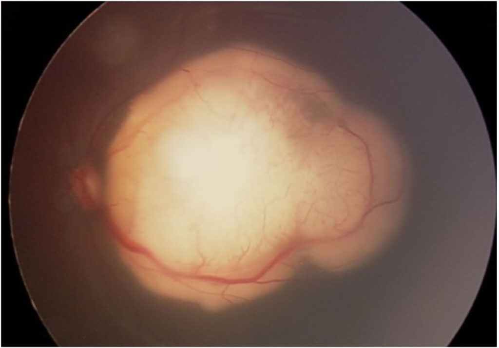
Retinoblastoma appears as a white or cream mass arising from the retina.
Dilated, tortuous feeder vessels enter the tumor itself.
In addition, chalky calcification, vitreous seeds and subretinal seeds point strongly to malignancy.
Finally, tumor cells may layer as a pseudohypopyon in the anterior chamber.
Vessel Pattern in Coats vs Retinoblastoma
In Coats, abnormal vessels leak into a detachment without a solid mass beneath them.
In retinoblastoma, however, dilated vessels feed and drain a solid tumor.
Therefore, trace each abnormal vessel to see whether it ends in exudate or in tissue.
Total Detachment in Coats vs Retinoblastoma
Advanced Coats disease can produce a bullous total detachment directly behind the lens.
Surface telangiectatic vessels then favor Coats.
Exophytic retinoblastoma, however, can also cause a total detachment that hides the mass (see exudative retinal detachment).
As a result, imaging decides most of these cases.
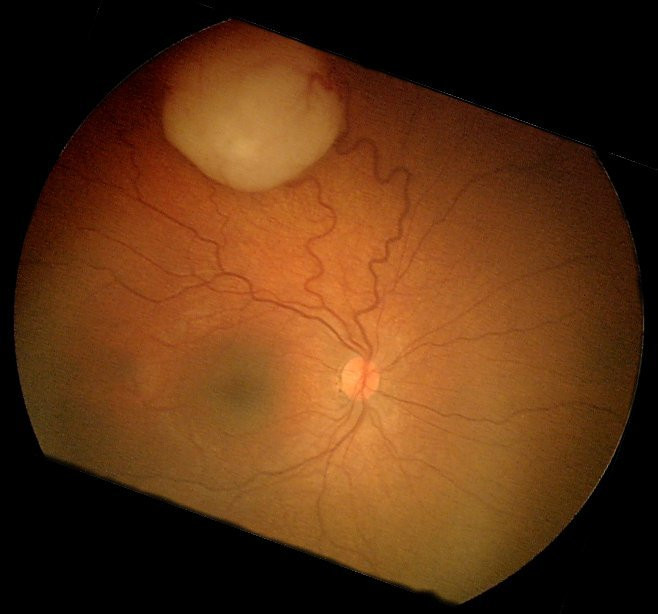
Ultrasound in Coats vs Retinoblastoma
B-scan ultrasound is the fastest and safest office test.
Retinoblastoma shows a solid mass with high internal reflectivity and calcific shadowing.
Coats disease, in contrast, shows detached retina over fluid with fine mobile echoes and no solid calcified mass.
However, longstanding Coats can rarely calcify, so read the echoes alongside the fundus picture.
Fluorescein Angiography
Widefield angiography, often under anesthesia, maps retinal vascular detail.
Coats disease shows telangiectasia, aneurysms, leakage and peripheral nonperfusion.
Retinoblastoma typically shows intrinsic tumor vessels and late hyperfluorescence of the mass.
Also, angiography guides laser to leaking vessels in Coats.
MRI in Coats vs Retinoblastoma
MRI is the preferred cross-sectional study when retinoblastoma remains possible.
It defines the mass, optic nerve invasion and extraocular spread.
Coats exudate, by contrast, shows fluid and lipid signal without an enhancing tumor.
However, avoid CT when possible, because radiation raises second cancer risk in germline RB1 carriers.
No Biopsy Before Imaging
Needle biopsy or vitrectomy can seed retinoblastoma cells outside the globe.
Therefore, avoid intraocular biopsy when a tumor remains on the list.
Also, drain subretinal fluid in suspected Coats only after imaging excludes a mass.
Examination Under Anesthesia
Most young children need an examination under anesthesia for complete assessment.
The surgeon then examines both fundi, takes widefield photos and repeats ultrasound.
Finally, the fellow eye always needs a full dilated examination.
Treatment After the Diagnosis
Coats treatment targets leaking vessels with laser photocoagulation or cryotherapy.
Anti-VEGF injections are sometimes added, although evidence remains limited.
Retinoblastoma, however, follows ocular oncology protocols with chemotherapy, focal therapy or enucleation.
Counseling Families
Parents fear cancer from the first mention of a white pupil.
Therefore, explain which findings support each diagnosis and which tests remain.
When retinoblastoma is confirmed, offer genetic counseling and RB1 testing for the family.
Pitfalls to Avoid
- Do not trust the reflex color alone
- Never biopsy or drain before imaging excludes a tumor
- Do not dismiss calcification as old Coats without careful review
- Remember diffuse infiltrating retinoblastoma in older children, which may lack obvious calcification
- Always examine the fellow eye


Document what you see
Two smartphone imaging tools built for everyday clinic use — one for the slit lamp, one for the fundus.
From Choroida — the team behind this siteReferences
- Shields JA, Shields CL, Honavar SG, Demirci H, Cater J. Classification and management of Coats disease: the 2000 Proctor Lecture. Am J Ophthalmol. 2001;131:572-583.
- Shields CL, Schoenberg E, Kocher K, et al. Lesions simulating retinoblastoma (pseudoretinoblastoma) in 604 cases: results based on age at presentation. Ophthalmology. 2013;120:311-316.
- Dimaras H, Kimani K, Dimba EA, et al. Retinoblastoma. Lancet. 2012;379:1436-1446.
- de Graaf P, Göricke S, Rodjan F, et al. Guidelines for imaging retinoblastoma: imaging principles and MRI standardization. Pediatr Radiol. 2012;42:2-14.
- Bowling B. Kanski’s Clinical Ophthalmology: A Systematic Approach. 8th ed. Elsevier; 2016.

Writes and reviews the clinical articles on Educate. Every post is checked against peer-reviewed sources before it goes live.