
CASE REPORT
Case report of Coats disease 25-year-old male patient presented with a gradual diminution of vision in his right eye, for the past 6 months. Ocular examination on presentation revealed a vision of 6/60 in his right eye with no improvement.

The vision in his left eye was normal i.e. 6/6. The intraocular pressure (IOP) of the right and left eye, measured using a non-contact tonometer (NCT), was 14 mm of Hg and 13 mm of Hg, respectively.
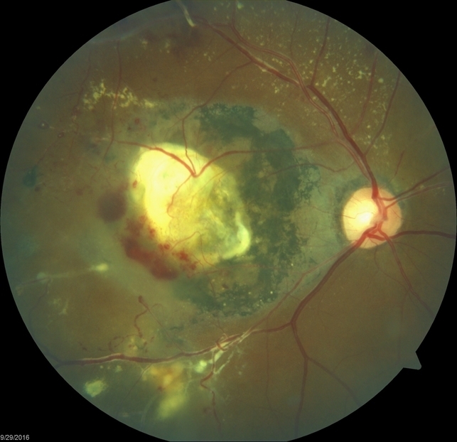
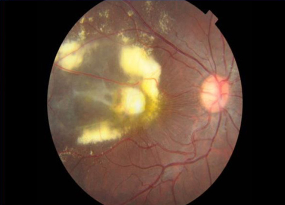
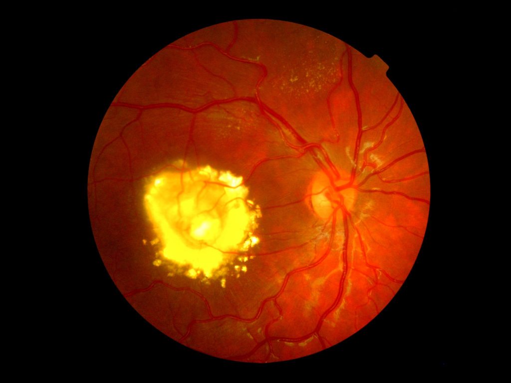
Slit-lamp examination revealed no specific findings in the anterior segment and no rubeosis. Dilated indirect fundoscopy revealed subretinal exudation extending into the fovea. Fundus Fluorescein Angiography (FFA) revealed abnormal inferotemporal vessels with aneurysmal dilatation and telangiectasia, which leaked profusely.
Peripheral to the abnormal vasculature was an area of capillary non-perfusion. Optical Coherence Tomography (OCT) done subsequently revealed cystoid macular edema. Left eye examination including fundoscopy, FFA, and OCT was all within normal limits (WNL). The patient was diagnosed as a case of Coats’ disease
DISEASE of Coats disease
It is a telangiectatic neovascular disease of the retina of unknown etiology that frequently affects unilateral eyes of young males.
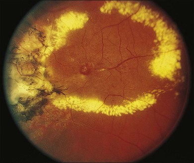
George Coats in 1908 described the histopathological features of enucleated eyes with massive exudation. He divided the morphology into 3 groups:
- Group I demonstrated massive subretinal exudate alone.
- Group II consisted of eyes with massive subretinal exudate, intra and subretinal hemorrhage, and retinal vascular dilatations.
- Group III included eyes with subretinal exudate and retinal arteriovenous malformations. Later, Von Hippel recognized group 3 as a separate entity called angiomatosis retinae.
A large study of 117 patients took the definition of Coats disease as ‘idiopathic retinal telangiectasia and intraretinal or subretinal exudation without appreciable signs of vitreoretinal traction.
MANAGEMENT of Coats disease
Observation- In minimal telangiectatic vessels with little or no exudation and no imminent threat to vision, or in painless and comfortable blind eyes
Laser treatment (low power, long duration, large spot burns to the telangiectatic vessels and aneurysms, also laser the peripheral capillary nonperfusion as per scatter laser settings) – in telangiectasia with exudation but no or minimal subretinal fluid
- Cryotherapy- for exudation with the subretinal fluid of such thickness that cryo reaction can reach the retina.
- Vitreoretinal surgery- for extensive retinal detachment not allowing cryo or laser.
- Enucleation- Painful blind eye or when retinoblastoma can not be ruled out by imaging and clinical examination.
- Anti-VEGF therapy- has been promising but the safety profile for children is as yet unclear.
- Intravitreal triamcinolone acetonide- may reduce the exudation, but risks of glaucoma and cataract are there.
would you have the interest to take retina images by smartphone?
Fundus photography is superior to fundus analysis as it enables intraocular pathologies to be photo captured and encrypted information to be shared with colleagues and patients.
Recent technologies allow smartphone-based attachments and integrated lens adaptors to transform the smartphone into a portable fundus camera, Slit lamp examination

REFERENCES
- Coats G. Forms of retinal diseases with massive exudation. R Lond Ophthalmol Hosp Rep. 1908;17:440-525
- Sigler EJ, Randolph JC, Calzada JI, Wilson MW, Haik BG. Current management of Coats disease. Surv Ophthalmol. 2014 Jan-Feb;59(1):30-46. doi: 10.1016/j.survophthal.2013.03.007. Epub 2013 Oct 15. Review. PubMed PMID:24138893.
- Shields JA, Shields CL, Honavar SG, Demirci H, Cater J. Classification and management of Coats disease: the 2000 Proctor Lecture. Am J Ophthalmol. 2001 May;131(5):572-83. PubMed PMID: 11336931.
- Shields JA, Shields CL, Honavar SG, Demirci H. Clinical variations and complications of Coats disease in 150 cases: the 2000 Sanford Gifford Memorial Lecture. Am J Ophthalmol. 2001 May;131(5):561-71. PubMed PMID: 11336930.
Read more: Retinal imaging by your smartphone, Bietti Crystalline Dystrophy, Choroidal Folds, Cherry red spot.
