AMD prognosis depends mainly on disease stage and on whether neovascularization develops.
Patients often ask one question first: will I go blind?
The answer is usually reassuring, because macular degeneration damages central vision and spares the peripheral field.
However, central loss can still remove reading, driving and face recognition.
What Patients Mean by “Blind”
Many patients picture total darkness when they hear the words macular degeneration.
AMD alone does not cause that outcome, because the peripheral retina keeps working.
However, some patients reach legal blindness from central loss in both eyes.
Therefore, separate the two meanings clearly in the first conversation.
AMD Prognosis by Beckman Stage
Early AMD with medium drusen carries a low short-term risk of advanced disease.
In contrast, intermediate AMD with large drusen or pigment changes carries a clearly higher risk.
Then advanced AMD, geographic atrophy or neovascular disease, defines the visual outcome (see the Beckman stages).
Fundus Explorer Pro
Photograph the retinal findings described here with the phone already in your pocket — 22 D optics and built-in illumination in one handheld unit.
From Choroida — the team behind this siteAREDS Scale and AMD Prognosis
The AREDS simplified scale counts risk factors in both eyes.
Each eye scores one point for large drusen and one for pigment abnormalities.
The 5-year risk of advanced AMD then rises from about 0.5% with no factors to about 50% with four.
Therefore, a quick fundus count gives a useful number for counseling (see AREDS simplified severity scale).
Fellow Eye Risk in AMD Prognosis
Advanced AMD in one eye raises the risk for the second eye considerably.
Therefore, patients should test each eye separately with an Amsler grid, because the good eye often masks new distortion.
New distortion or a gray spot needs review within days, because early anti-VEGF treatment protects vision.
Dry AMD Prognosis
Dry AMD usually progresses slowly over years.
Many patients with early or intermediate disease keep good reading vision for a long time.
However, geographic atrophy enlarges steadily and gradually erodes the central field.
Vision often stays good while the fovea escapes, then falls once atrophy reaches the center.
Also, acuity alone can underestimate disability, because reading speed and dim light vision suffer first.
Wet AMD Prognosis
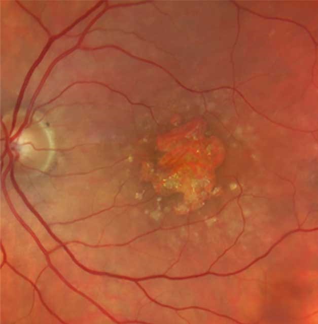
Untreated neovascular AMD often causes rapid central loss and ends in a disciform scar.
With monthly ranibizumab in MARINA, however, most eyes avoided moderate vision loss over two years.
Also, outcomes depend on early detection and on regular injections.
Long-Term Outcomes After Anti-VEGF
Long-term follow-up tempers the early trial results.
For example, in SEVEN-UP many treated eyes had lost vision about seven years later, often with less intensive treatment.
Macular atrophy and fibrosis also develop in some treated eyes.
As a result, explain that treatment controls the disease and does not cure it.
Signs That Worsen AMD Prognosis
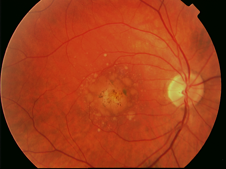
- Large drusen in both eyes
- Pigment clumping or hypopigmentation
- Reticular pseudodrusen
- Advanced AMD in the fellow eye
- Late presentation of neovascular AMD with poor acuity
- Subretinal fibrosis or foveal atrophy
OCT Findings That Refine AMD Prognosis
OCT adds information that the fundus view can miss.
For example, intraretinal hyperreflective foci and incomplete atrophy predict progression toward geographic atrophy.
Also, persistent intraretinal fluid in wet AMD often predicts poorer visual results.
Can Patients Change Their AMD Prognosis?
Some risk factors are modifiable.
First, smoking cessation lowers risk, and patients should hear this plainly.
In addition, AREDS supplements lowered progression to advanced AMD by about 25% over five years in higher-risk patients.
However, supplements show no clear benefit in early AMD.
Treatment for Geographic Atrophy
Pegcetacoplan and avacincaptad pegol, two complement inhibitors, slow the growth of geographic atrophy.
However, their trials did not show a clear benefit for visual acuity.
They also need frequent injections and carry risks, including conversion to neovascular AMD.
Therefore, present them as a way to slow structural loss, without promising better vision.
How to Explain AMD Prognosis to Patients
Start with the stage and type, then give a realistic range of outcomes.
Use plain words: central vision may fade, while side vision usually stays.
Next, explain warning symptoms and the follow-up plan.
Finally, offer low vision support early (see low vision rehabilitation in AMD).
Charles Bonnet Syndrome
Some patients with severe central loss see vivid images that are not there.
They often fear dementia and hide the symptom.
Therefore, ask directly and explain that these hallucinations reflect vision loss.
Pitfalls to Avoid
- Do not say blind without explaining central versus total loss
- Never judge geographic atrophy by acuity alone
- Do not skip the fellow eye at each visit
- Avoid promising vision gain from complement inhibitors


Document what you see
Two smartphone imaging tools built for everyday clinic use — one for the slit lamp, one for the fundus.
From Choroida — the team behind this siteReferences
- Ferris FL 3rd, Wilkinson CP, Bird A, et al. Clinical classification of age-related macular degeneration. Ophthalmology. 2013;120:844-851.
- Ferris FL, Davis MD, Clemons TE, et al. A simplified severity scale for age-related macular degeneration: AREDS report no. 18. Arch Ophthalmol. 2005;123:1570-1574.
- Age-Related Eye Disease Study Research Group. A randomized, placebo-controlled, clinical trial of high-dose supplementation with vitamins C and E, beta carotene, and zinc for age-related macular degeneration and vision loss: AREDS report no. 8. Arch Ophthalmol. 2001;119:1417-1436.
- Rosenfeld PJ, Brown DM, Heier JS, et al. Ranibizumab for neovascular age-related macular degeneration. N Engl J Med. 2006;355:1419-1431.
- Rofagha S, Bhisitkul RB, Boyer DS, et al. Seven-year outcomes in ranibizumab-treated patients in ANCHOR, MARINA, and HORIZON: a multicenter cohort study (SEVEN-UP). Ophthalmology. 2013;120:2292-2299.

Writes and reviews the clinical articles on Educate. Every post is checked against peer-reviewed sources before it goes live.