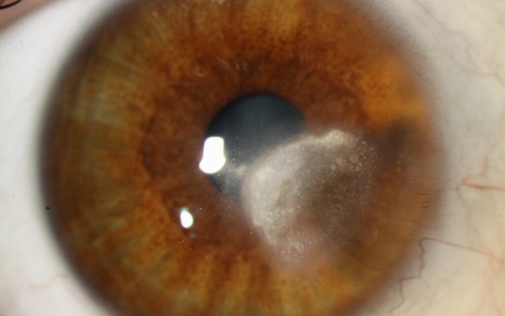
Lipid keratopathy is the deposition of cholesterol, fatty acids and phospholipids within the corneal stroma.
It appears at the slit lamp as a dense yellow-white opacity, often with feathery edges.
Most cases are secondary and follow corneal neovascularization.
Therefore, the examiner should search for a feeding vessel first.
What Lipid Keratopathy Looks Like
The opacity often takes a fan or disc shape, with a feathery margin.
In secondary disease, a vessel commonly runs toward the deposit.
Also, fine cholesterol crystals may glitter within denser areas.
Primary Lipid Keratopathy
Primary disease is rare and appears without previous corneal injury or inflammation.
Slit lamp examination shows no obvious vessels, and serum lipids are typically normal.
However, search carefully before using this label because regressed vessels are easy to miss.
All-fit Slit-Lamp Adapter
Record and share exactly what you see at the slit lamp. One adapter fits any slit lamp or surgical microscope — and any smartphone.
From Choroida — the team behind this siteSecondary Lipid Keratopathy
Secondary disease develops in a cornea that has already vascularized.
Common triggers include herpes simplex keratitis, herpes zoster keratitis and trauma.
In addition, interstitial keratitis, contact lens hypoxia and previous ulcers can precede it.
The inciting event may be years old, so a careful history often supplies the missing link.
Why Corneal Vessels Leave Lipid Behind
New corneal vessels are immature and more permeable than normal limbal vessels.
As a result, lipid-rich plasma can leak into the avascular stroma.
Clearance from this tissue is slow, so material accumulates around the vessel.
Symptoms of Lipid Keratopathy
Peripheral deposits may cause no symptoms and may be found on routine examination.
However, central deposits can reduce vision through opacity, glare and irregular astigmatism.
Pain and redness suggest active inflammation, because lipid itself is usually quiet.
Differential Diagnosis of Lipid Keratopathy
Several corneal opacities can mimic this finding, so location and associated signs guide the diagnosis.
- Arcus senilis: a peripheral ring with a clear zone beside the limbus
- Schnyder corneal dystrophy: bilateral inherited central haze or crystals
- Infectious crystalline keratopathy: branching deposits, often after grafting or steroid use
- Band keratopathy: interpalpebral calcium at the Bowman layer level
- Active stromal keratitis: infiltrate with edema and inflammation
Testing in Lipid Keratopathy
Slit lamp examination usually establishes the diagnosis.
Anterior segment OCT then measures deposit depth, which helps with surgical planning.
In addition, consider a fasting lipid profile in primary or bilateral disease.
Examination Pearls in Lipid Keratopathy
First, use a narrow slit beam to judge how deep the deposit sits in the stroma.
Then, try retroillumination, because it reveals fine ghost vessels that direct light hides.
Also, apply fluorescein to check whether the overlying epithelium is intact.
As a result, you can separate a quiet scar from active disease.
Treating the Cause First
Treatment starts with the disease that drives neovascularization.
For example, herpetic stromal disease may need antiviral therapy with carefully supervised topical steroids.
Similarly, contact lens hypoxia calls for a change in lens wear.
Without this control, new vessels may keep supplying lipid after any local procedure.
Medical Therapy for Lipid Keratopathy
Topical steroids reduce inflammation and may limit active vessel growth.
However, mature vessels respond less well, and established deposits may change little.
Steroids can also raise intraocular pressure and promote cataract, so monitor patients on prolonged courses.
Anti-VEGF Treatment
Subconjunctival or intracorneal bevacizumab targets the vascular supply, and its use here is off-label.
Oh and colleagues reported vessel regression and some lipid reduction in a small case series.
However, evidence is limited, and vessels can recur, so repeat treatment may be needed.
Vessel Occlusion Procedures
Argon or yellow dye laser and fine needle diathermy can close selected feeder vessels.
Once the supply closes, deposits may slowly regress over months.
However, these procedures may cause hemorrhage, inflammation or corneal thinning, and vessels can reopen.
Surgery for Lipid Keratopathy
Lamellar or penetrating keratoplasty may be considered when central scarring limits vision.
Because vascularized host beds raise rejection risk, surgeons often control vessels beforehand.
Surgery removes the opacity, while the underlying disease can still recur.
Counseling Patients
First, explain that the deposit follows leaky corneal vessels and usually reflects past corneal disease.
Next, tell them that treatment aims to stop progression and seldom clears the deposit completely.
Therefore, set realistic visual goals before any procedure.
In addition, advise urgent review for pain, redness or a sudden drop in vision.
Documenting Progress
For example, serial slit lamp photographs allow objective comparison of deposit size and vessel caliber.
Also, anterior segment OCT can show whether the depth has changed between visits.
Follow-Up in Lipid Keratopathy
Peripheral, stable deposits with quiet vessels may need observation alone.
Then, record acuity, deposit margins and vessel activity at each visit.
Finally, tell patients that complete clearing is uncommon and that new pain or redness needs prompt review.
Pitfalls to Avoid
- Do not call the disease primary before excluding subtle vessels
- Never start steroids for a painful cornea without excluding infection
- Do not promise complete clearing after vessel closure


Document what you see
Two smartphone imaging tools built for everyday clinic use — one for the slit lamp, one for the fundus.
From Choroida — the team behind this siteReferences
- Hall MN, Moshirfar M, Amin-Javaheri A, et al. Lipid keratopathy: a review of pathophysiology, differential diagnosis, and management. Ophthalmol Ther. 2020;9:833-852.
- Cogan DG, Kuwabara T. Lipid keratopathy and atheroma. Circulation. 1958;18:519-525.
- Oh JY, Kim MK, Wee WR. Subconjunctival and intracorneal bevacizumab injection for corneal neovascularization in lipid keratopathy. Cornea. 2009;28:1070-1073.
- Pillai CT, Dua HS, Hossain P. Fine needle diathermy occlusion of corneal vessels. Invest Ophthalmol Vis Sci. 2000;41:2148-2153.

Writes and reviews the clinical articles on Educate. Every post is checked against peer-reviewed sources before it goes live.