Roper-Hall classification sorts acute ocular burns into four grades using corneal clarity and limbal ischemia.
Roper-Hall published the system in 1965, building on the earlier Hughes and Ballen schemes.
Clinicians still use it because it needs only a slit lamp and a careful look.
What the Roper-Hall Classification Measures
The system combines two findings that reflect different types of tissue damage.
First, corneal haze suggests how far the injury has extended into the stroma.
Second, limbal ischemia reflects damage to the limbal vessels and, by inference, the stem cell niche.
Because corneal resurfacing depends on these stem cells, the limbal finding carries strong prognostic weight.
The Four Grades in the Roper-Hall Classification
- Grade I: corneal epithelial damage, no limbal ischemia, good prognosis
- In Grade II, the cornea is hazy but iris detail is visible, ischemia involves under one third of the limbus, and prognosis is good
- With Grade III, total epithelial loss and stromal haze obscure iris detail, one third to one half of the limbus is ischemic, and prognosis is guarded
- Grade IV: opaque cornea, obscured iris and pupil, ischemia over one half of the limbus, poor prognosis
All-fit Slit-Lamp Adapter
Record and share exactly what you see at the slit lamp. One adapter fits any slit lamp or surgical microscope — and any smartphone.
From Choroida — the team behind this siteGrades I and II at the Slit Lamp
Grade I shows an epithelial defect or punctate staining with a clear stroma.
Limbal vessels fill normally, so stem cell reserve should remain adequate.
In Grade II, haze appears, yet the iris stays visible.
Healthy adjacent limbus can usually repopulate the injured sector, although some eyes develop localized scarring.
Grades III and IV at the Slit Lamp
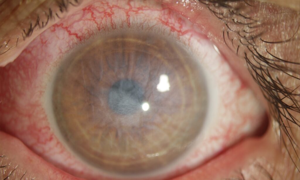
In Grade III, denser haze hides the iris, and healing often takes longer.
Persistent epithelial defects therefore become more likely.
Grade IV eyes look porcelain white, and the conjunctiva often appears devitalized.
As a result, clinicians expect delayed healing, stromal melting, vascularization and severe surface failure.
Examining the Eye Before Grading
Irrigation comes first, and grading should never delay it.
Next, confirm that the conjunctival pH has normalized and sweep the fornices for retained particles.
Then use fluorescein to map the epithelial defect, including the conjunctiva.
Finally, examine each limbal sector for blanching and absent vascular filling.
Limits of the Roper-Hall Classification
The system does not score conjunctival involvement, although conjunctiva contributes to surface recovery.
In addition, every eye with more than half of the limbus involved falls into Grade IV.
Corneal edema can also make early haze hard to interpret.
Therefore, repeat the grading at follow-up and record each result with its date.
Roper-Hall Classification Versus Dua Grading
Dua and colleagues proposed a six-grade system in 2001.
It records limbal involvement in clock hours and conjunctival involvement as a percentage.
Gupta and colleagues later found that the Dua system predicted outcome better in severe burns.
However, the two systems performed similarly in milder injuries.
Treatment Planning With the Roper-Hall Classification
The grade guides the intensity of care, but it does not prescribe one fixed regimen.
Mild burns usually receive preservative-free lubricants, a topical antibiotic and limited topical steroid.
Severe burns may also need cycloplegia, pressure control, and adjuncts such as ascorbate or doxycycline.
Steroids reduce inflammation, yet prolonged use can increase melting risk once stromal repair begins.
Amniotic Membrane by Grade
Amniotic membrane supports epithelial healing and may reduce surface inflammation.
In a randomized trial, Tandon and colleagues reported faster epithelial healing in moderate burns.
However, they found no clear benefit in severe burns.
It cannot replace a destroyed stem cell population, so counsel patients with Grade IV injury accordingly.
What Severe Grades Predict Later
Higher grades raise concern for limbal stem cell deficiency, conjunctivalization and vascularized scarring.
Symblepharon, secondary glaucoma, cataract and perforation may also follow.
Glaucoma can develop silently, while an opaque cornea complicates pressure measurement.
Limbal stem cell transplantation may rebuild the surface later, but keratoplasty alone tends to fail on a deficient surface.
Counseling Patients by Grade
Give the grade as an early estimate, and explain that it may change.
Patients with Grade I or II injury can usually expect good recovery, although they need follow-up.
For Grade III or IV injury, discuss prolonged treatment, possible surgery and uncertain vision.
Pitfalls to Avoid
- Do not delay irrigation to assign a grade
- Do not read a white, quiet limbus as a mild injury
- Avoid treating the first grade as final
- Do not ignore conjunctival loss because the grade omits it
- Never stop monitoring pressure after the epithelium heals


Document what you see
Two smartphone imaging tools built for everyday clinic use — one for the slit lamp, one for the fundus.
From Choroida — the team behind this siteReferences
- Roper-Hall MJ. Thermal and chemical burns. Trans Ophthalmol Soc U K. 1965;85:631-653.
- Dua HS, King AJ, Joseph A. A new classification of ocular surface burns. Br J Ophthalmol. 2001;85:1379-1383.
- Gupta N, Kalaivani M, Tandon R. Comparison of prognostic value of Roper Hall and Dua classification systems in acute ocular burns. Br J Ophthalmol. 2011;95:194-198.
- Tandon R, Gupta N, Kalaivani M, et al. Amniotic membrane transplantation as an adjunct to medical therapy in acute ocular burns. Br J Ophthalmol. 2011;95:199-204.
- Sharma N, Kaur M, Agarwal T, et al. Treatment of acute ocular chemical burns. Surv Ophthalmol. 2018;63:214-235.

Writes and reviews the clinical articles on Educate. Every post is checked against peer-reviewed sources before it goes live.