IVTS classification is an OCT-based system that describes the vitreomacular interface in anatomical terms.
The International Vitreomacular Traction Study Group published it in 2013, and it sorts eyes into vitreomacular adhesion, vitreomacular traction and full-thickness macular hole. As a result, clinicians can report scans consistently.
Why the IVTS Classification Was Developed
Gass built his macular hole stages on biomicroscopy, before OCT showed the interface in detail. However, his stages can imply steady progression, while many traction eyes remain stable or release spontaneously. Therefore, the IVTS group focused on the anatomy visible on the scan.
How to Read the OCT
Review the full volume, because one line scan can miss a small hole. First, trace the posterior hyaloid toward the fovea, then inspect the foveal contour and the outer retinal layers, and finally measure the attachment or hole.
Fundus Explorer Pro
Photograph the retinal findings described here with the phone already in your pocket — 22 D optics and built-in illumination in one handheld unit.
From Choroida — the team behind this siteVitreomacular Adhesion in IVTS Classification
Vitreomacular adhesion means the perifoveal vitreous has separated, while the cortex stays attached within 3 mm of the fovea, and the foveal contour and underlying retina remain unchanged on OCT. Because the retina looks undisturbed, adhesion alone rarely explains visual symptoms (see vitreomacular adhesion), and therefore observation usually suffices.
Vitreomacular Traction on OCT
Vitreomacular traction shares the same attachment pattern, but the retina now shows structural change. Findings may include foveal distortion, pseudocysts, schisis or subretinal fluid, but the retinal layers are not interrupted through their full thickness. Patients may notice metamorphopsia or blur (see vitreomacular traction).
Focal and Broad Attachment
The system also grades the width of the vitreous attachment. Focal attachment measures 1500 microns or less, while broad attachment exceeds 1500 microns and may also coexist with epiretinal membrane.
Isolated and Concurrent Disease
Adhesion or traction is isolated when no other macular disease appears on the scan, and it is concurrent when conditions such as epiretinal membrane or macular degeneration coexist. For example, vitrectomy cannot reverse vision loss caused by advanced macular atrophy.
Full-Thickness Holes in IVTS Classification
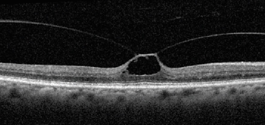
A full-thickness macular hole interrupts all retinal layers from the internal limiting membrane to the RPE, and clinicians then record whether vitreomacular traction persists. They also separate primary holes from secondary holes, for example traumatic macular hole or high myopia.
Hole Size Categories
IVTS classification measures the aperture at its narrowest point, with small holes measuring 250 microns or less. Medium holes measure up to 400 microns, while large holes exceed 400 microns, and these groups help with surgical counseling (see macular hole staging).
Mapping Gass Stages to IVTS Classification
Gass stage 1 corresponds to vitreomacular traction, and next, stage 2 corresponds to a small or medium hole with traction. Stage 3 corresponds to a medium or large hole with traction, and finally, stage 4 corresponds to a hole without traction.
Lamellar Holes and Pseudoholes
A 2020 consensus later defined lamellar macular hole using OCT criteria. Therefore, confirm whether all retinal layers are interrupted before assigning a diagnosis (see lamellar versus full-thickness hole).
Example 1: Incidental Adhesion
A patient has no complaint, and OCT shows an 800-micron attachment with a normal foveal contour. The label is focal, isolated vitreomacular adhesion, and therefore the clinician can reassure the patient.
Example 2: Symptomatic Traction
A patient reports metamorphopsia, and OCT shows a 600-micron attachment lifting the fovea. Pseudocysts are present, while the retinal layers remain uninterrupted, so the label is focal, isolated vitreomacular traction.
Example 3: Primary Macular Hole
A patient has a central scotoma, and OCT shows all layers interrupted, while the narrowest aperture measures 320 microns and the hyaloid remains attached. The label is a medium, primary full-thickness hole with traction, so the clinician should discuss vitrectomy.
Treatment Choices After IVTS Classification
Observation suits many mild traction cases, because spontaneous release can occur. Ocriplasmin improved traction resolution over placebo at day 28 in MIVI-TRUST, 26.5% versus 10.1%, although availability varies. Vitrectomy directly relieves traction and treats holes, but it carries risks including cataract and retinal detachment, and it also cannot guarantee normal vision.
Follow-Up and Limits of IVTS Classification
Repeat OCT when symptoms change, because traction can release or a hole can develop. However, the system does not grade symptoms or visual potential.
Pitfalls to Avoid
- Do not diagnose a hole from a single off-center scan
- Do not call adhesion pathologic when the fovea looks normal
- Never assume that every traction eye will progress to a hole


Document what you see
Two smartphone imaging tools built for everyday clinic use — one for the slit lamp, one for the fundus.
From Choroida — the team behind this siteReferences
- Duker JS, Kaiser PK, Binder S, et al. The International Vitreomacular Traction Study Group classification of vitreomacular adhesion, traction, and macular hole. Ophthalmology. 2013;120:2611-2619.
- Gass JDM. Reappraisal of biomicroscopic classification of stages of development of a macular hole. Am J Ophthalmol. 1995;119:752-759.
- Stalmans P, Benz MS, Gandorfer A, et al. Enzymatic vitreolysis with ocriplasmin for vitreomacular traction and macular holes. N Engl J Med. 2012;367:606-615.
- Hubschman JP, Govetto A, Spaide RF, et al. Optical coherence tomography-based consensus definition for lamellar macular hole. Br J Ophthalmol. 2020;104:1741-1747.

Writes and reviews the clinical articles on Educate. Every post is checked against peer-reviewed sources before it goes live.