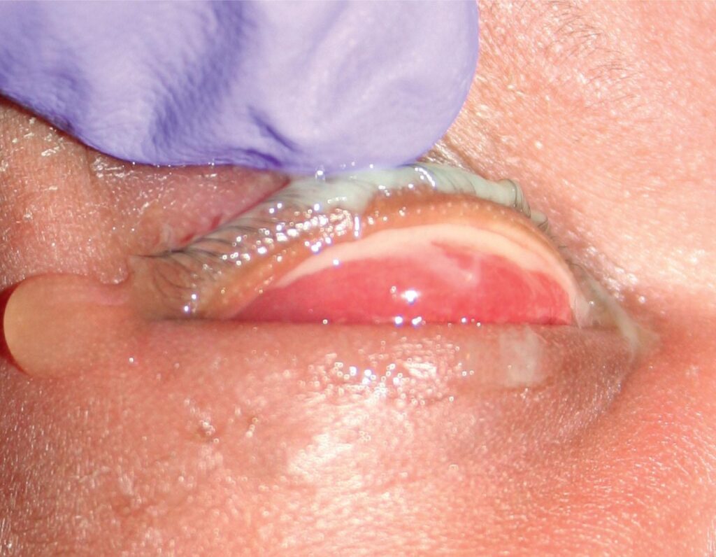
Pseudomembranous conjunctivitis is conjunctival inflammation with a fibrin coat over the tarsal surface.
The finding narrows the differential because only a few causes produce it, and it also predicts scarring, so it deserves careful lid eversion.
What a Pseudomembrane Is
A pseudomembrane forms when fibrin and inflammatory cells leak from inflamed vessels, and the exudate coagulates on the epithelial surface but does not invade it.
As a result, it peels away easily and the bed stays smooth, although slight oozing may follow and brisk bleeding is unusual.
True Membranes Versus Pseudomembranes
A true membrane integrates with the superficial epithelium. Therefore, stripping it tears the tissue and leaves a raw, bleeding surface.
In practice, the two overlap, and adherence depends partly on the age of the lesion. Both signal severe inflammation, so the cause matters more than the label.
All-fit Slit-Lamp Adapter
Record and share exactly what you see at the slit lamp. One adapter fits any slit lamp or surgical microscope — and any smartphone.
From Choroida — the team behind this siteExam Signs of Pseudomembranous Conjunctivitis
Evert both upper lids, because membranes often hide on the tarsal plate. Look for a gray or yellow-white sheet that follows the tarsal contour.
Then touch it gently with a moistened swab to test adherence, and also record chemosis, follicles, discharge, preauricular nodes and corneal staining.
Adenovirus as the Leading Cause
Epidemic keratoconjunctivitis is the commonest cause seen in clinic, and serotypes such as 8, 19 and 37 produce the most severe forms.
Patients report a watery, gritty eye that started on one side and spread to the other. Furthermore, membranes accompany more severe disease with a higher chance of lasting fibrosis (see adenoviral infection control).
Bacterial Causes of Pseudomembranous Conjunctivitis
Neisseria gonorrhoeae causes hyperacute purulent conjunctivitis that may form membranes, and copious discharge, lid swelling and rapid onset point toward it.
In addition, it can melt the cornea within days (see gonococcal conjunctivitis). Streptococcus pyogenes and Corynebacterium diphtheriae classically form true membranes.
Stevens-Johnson Syndrome and Toxic Epidermal Necrolysis
SJS and TEN can strip the conjunctival epithelium within days of a drug reaction, and pseudomembranes then cover the lids and fornices.
Because the damage progresses quickly, early ophthalmic review is essential. Many centers place amniotic membrane early to limit later cicatrization.
Chemical Injury and Pseudomembranous Conjunctivitis
Alkali and acid burns can produce fibrinous membranes during the acute phase. First, irrigate until the pH normalizes, then examine for limbal ischemia. Membranes here forecast symblepharon, so daily fornix checks are worthwhile.
Other Causes to Consider
- Primary herpes simplex infection in children
- Neonatal chlamydial conjunctivitis
- Ligneous conjunctivitis from plasminogen deficiency (see ligneous conjunctivitis)
- Mucous membrane pemphigoid during an active flare
- Ocular graft-versus-host disease
- Retained foreign bodies and severe toxic reactions
History That Narrows the Cause
Ask about sick contacts, recent eye clinic visits and the time course, and also ask about new drugs, sexual exposures and chemical splashes. A child with skin vesicles suggests herpes, while recurrent woody membranes suggest ligneous disease.
Tests in Pseudomembranous Conjunctivitis
Most adenoviral cases need only a clinical diagnosis. However, a rapid antigen test or PCR helps when the picture is unclear.
Send Gram stain and culture for purulent or hyperacute disease, while recurrent or atypical membranes justify plasminogen testing or biopsy.
Should You Peel the Membrane?
Peeling is common practice, although controlled trial evidence is limited. Removal often eases foreign body sensation, lets topical drugs reach the surface, and may also reduce the risk of fibrosis and symblepharon. On the other hand, membranes often re-form, so repeat removal every few days may be needed.
How to Peel Safely
Instill topical anesthetic and use a cotton-tipped applicator or fine forceps, then lift the edge and roll the sheet off the tarsus. Stop if the tissue bleeds briskly, because that suggests a true membrane.
Medical Treatment of Pseudomembranous Conjunctivitis
Treat the cause first, with systemic ceftriaxone for gonococcal disease. For adenoviral membranes, many clinicians add a short course of topical steroid.
Steroids reduce inflammation and membrane formation; however, they may prolong viral shedding. Therefore, taper under supervision and monitor intraocular pressure.
Scarring Risk in Pseudomembranous Conjunctivitis
Membranes damage the basement membrane and expose the substantia propria. As a result, fibroblasts lay down scar that can fuse opposing surfaces.
Complications include subepithelial fibrosis, symblepharon, fornix shortening and trichiasis. In addition, goblet cell loss can leave chronic dry eye.
Counseling the Patient
Explain that the white layer is inflammatory material and not pus or tissue, and warn that it may return and need more than one peel. Adenoviral cases are contagious, so advise hand hygiene and time off work.
Follow-Up Visits
Review severe cases every few days during the acute phase. At each visit, evert the lids and inspect the fornices for early symblepharon. Later, check for subepithelial infiltrates and residual tear film disturbance.
Pitfalls to Avoid
- Do not skip lid eversion in a red, swollen eye
- Never start steroids before excluding herpes simplex
- Do not miss hyperacute discharge that suggests gonococcus
- Do not ignore recurrent membranes, because ligneous disease needs systemic workup


Document what you see
Two smartphone imaging tools built for everyday clinic use — one for the slit lamp, one for the fundus.
From Choroida — the team behind this siteReferences
- Varu DM, Rhee MK, Akpek EK, et al. Conjunctivitis Preferred Practice Pattern. Ophthalmology. 2019;126:P94-P169.
- Azari AA, Barney NP. Conjunctivitis: a systematic review of diagnosis and treatment. JAMA. 2013;310:1721-1729.
- Jhanji V, Chan TC, Li EY, Agarwal K, Vajpayee RB. Adenoviral keratoconjunctivitis. Surv Ophthalmol. 2015;60:435-443.
- Bowling B. Kanski’s Clinical Ophthalmology: A Systematic Approach. 8th ed. Elsevier. 2016.

Writes and reviews the clinical articles on Educate. Every post is checked against peer-reviewed sources before it goes live.