Solar retinopathy recovery usually unfolds over weeks to months, and most eyes regain useful central vision.
The injury follows direct viewing of the sun, most often during a solar eclipse. However, a small foveal defect and a faint scotoma can last for many years.
Mechanism Behind the Injury
Short-wavelength visible light triggers a photochemical reaction in the photoreceptors and retinal pigment epithelium, while thermal damage plays a smaller role at typical viewing durations. As a result, the fovea shows outer retinal disruption with little inflammation, and long viewing time, clear young lenses and photosensitizing drugs all increase the light dose.
Symptoms in the First Days
Patients notice blurred central vision, a central or paracentral scotoma, and sometimes metamorphopsia, and symptoms often start within hours, so the history usually links them to the exposure. Some also report chromatopsia, afterimages or headache, and because both eyes usually viewed the sun, the damage is often bilateral and asymmetric.
Fundus Explorer Pro
Photograph the retinal findings described here with the phone already in your pocket — 22 D optics and built-in illumination in one handheld unit.
From Choroida — the team behind this siteAcute Fundus Findings
Early on, the examiner may see a small yellow-white foveal spot, although the fundus can also look entirely normal, which delays diagnosis.
Over the following days to weeks, the spot fades into a reddish, sharply outlined foveal lesion. For a broad review of the condition, see solar retinopathy: an in-depth look.
OCT Signs of Solar Retinopathy Recovery
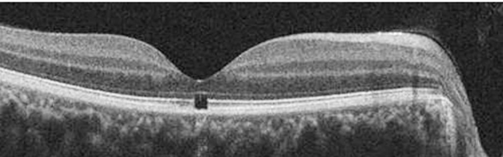
Acute OCT typically shows foveal hyperreflectivity spanning the outer retinal layers, which next resolves and leaves a focal hyporeflective gap at the ellipsoid and interdigitation zones.
The gap often looks like a small box at the foveal center. Therefore, OCT confirms the diagnosis when the fundus looks subtle.
Visual Acuity During Solar Retinopathy Recovery
Presenting acuity ranges from near normal to moderate loss, and most published series describe gradual improvement over the first several months.
Many patients return to 20/20 or near it, while a minority stabilize with mild reduction, and slow gains may continue for up to a year in some reports.
First Month of Solar Retinopathy Recovery
During the first weeks, symptoms usually change little, while the acute OCT hyperreflectivity begins to fade. The patient may also notice the scotoma shrinking or becoming less dense, while you record acuity and capture a baseline OCT for later comparison.
Three to Six Months
This window brings the largest functional gains in most eyes, as metamorphopsia often eases and reading speed improves. However, the outer retinal defect on OCT may shrink without closing completely, so structure and function therefore do not always move together.
What Persists Long Term
A small central or paracentral scotoma persists in some patients, and many describe, for example, a fixed dot that moves with gaze. In addition, a lasting foveal ellipsoid zone defect is common even with 20/20 acuity, and faint metamorphopsia on Amsler grid testing may also remain.
What Determines Solar Retinopathy Recovery
Prognosis depends mainly on the dose of light reaching the fovea, and reported factors that may worsen the outlook include:
- Longer viewing time and repeated exposures
- Wider outer retinal defect on initial OCT
- Worse presenting visual acuity
- Young age with clear crystalline lenses
- Viewing through improper filters or optical devices
Treatment Has Limited Evidence
No treatment has proven benefit in solar retinopathy, although some clinicians prescribe oral corticosteroids early, based on case reports. However, no controlled trial supports this, and steroids carry systemic risks, so observation is the standard approach for most patients.
Counseling on Solar Retinopathy Recovery
Start with honest expectations: improvement is likely but often incomplete. Explain that vision tends to improve slowly over months, and warn that a small blind spot may stay and that most people adapt to it. Then advise strict sun avoidance, because repeat exposure can add damage.
Follow-Up During Solar Retinopathy Recovery
First, review the patient at about one month, then at three and six months, and at each visit check acuity, Amsler grid and OCT. Microperimetry, when available, maps the scotoma and tracks its density. Discharge once findings stabilize, while advising a prompt return for new symptoms.
Differential Diagnosis to Exclude
A foveal outer retinal defect has several mimics.
- Lamellar or full-thickness macular hole
- Laser pointer maculopathy
- Macular telangiectasia type 2
- Tamoxifen or other drug-related maculopathy
- Whiplash maculopathy after blunt trauma
A full-thickness hole needs surgery and follows a different course (see macular hole recovery), so a careful exposure history and OCT usually settle the question.
Prevention for Future Eclipses
Advise only certified solar filters that meet the ISO 12312-2 standard. Similarly, sunglasses, smoked glass and exposed film do not protect the retina, and patients should never view the sun through binoculars or telescopes without proper front filters.
Pitfalls in Solar Retinopathy Recovery
- Do not dismiss symptoms because the fundus looks normal
- Avoid promising full recovery at the first visit
- Do not overlook the fellow eye
- Avoid unproven treatment without discussing risks
- Do not stop follow-up before OCT stabilizes


Document what you see
Two smartphone imaging tools built for everyday clinic use — one for the slit lamp, one for the fundus.
From Choroida — the team behind this siteReferences
- Wong SC, Eke T, Ziakas NG. Eclipse burns: a prospective study of solar retinopathy following the 1999 solar eclipse. Lancet. 2001;357:199-200.
- Wu CY, Jansen ME, Andrade J, et al. Acute solar retinopathy imaged with adaptive optics, optical coherence tomography angiography, and en face optical coherence tomography. JAMA Ophthalmol. 2018;136:82-85.
- Jain A, Desai RU, Charalel RA, et al. Solar retinopathy: comparison of optical coherence tomography (OCT) and fluorescein angiography (FA). Retina. 2009;29:1340-1345.
- Mainster MA, Turner PL. Retinal injuries from light: mechanisms, hazards, and prevention. In: Ryan SJ, ed. Retina. 5th ed. Elsevier; 2013.
- Bowling B. Kanski’s Clinical Ophthalmology: A Systematic Approach. 8th ed. Elsevier; 2016.
Test yourself
A few questions straight from this article.
-
What is the main mechanism of retinal injury in solar retinopathy?
Short-wavelength visible light triggers a photochemical reaction in photoreceptors and RPE, with thermal damage playing a smaller role at typical viewing durations. -
What laterality pattern is typical of solar retinopathy after eclipse viewing?
Because both eyes usually viewed the sun, the damage is often bilateral and asymmetric, so the fellow eye must not be overlooked. -
What may the fundus show early in acute solar retinopathy?
Early on a small yellow-white foveal spot may be seen, though the fundus can look entirely normal; it later fades into a reddish, sharply outlined lesion. -
What OCT finding persists after the acute hyperreflectivity of solar retinopathy resolves?
Acute outer retinal hyperreflectivity resolves to leave a focal, often box-like hyporeflective gap at the ellipsoid and interdigitation zones. -
In solar retinopathy, which period usually brings the largest functional gains?
The three-to-six-month window brings the largest functional gains in most eyes, as metamorphopsia eases and reading speed improves. -
Which long-term structural finding is common after solar retinopathy, even with 20/20 acuity?
A lasting foveal ellipsoid zone defect is common even with 20/20 acuity, and a small scotoma or faint metamorphopsia may remain. -
Which initial finding is reported to worsen the visual outlook in solar retinopathy?
Prognosis depends mainly on the light dose; a wider initial OCT defect, worse presenting acuity, longer or repeated viewing and young clear lenses may worsen the outlook. -
What is the standard management approach for most patients with solar retinopathy?
No treatment has proven benefit; some give early oral corticosteroids on case-report evidence, but no controlled trial supports this, so observation is standard. -
Which eye protection should be advised for viewing a future solar eclipse?
Only certified solar filters meeting ISO 12312-2 should be used; sunglasses, smoked glass and exposed film do not protect the retina. -
What follow-up schedule is suggested during solar retinopathy recovery?
Review at about one month, then three and six months, checking acuity, Amsler grid and OCT, and discharge once findings stabilise.

Writes and reviews the clinical articles on Educate. Every post is checked against peer-reviewed sources before it goes live.