RVO stroke risk is a frequent patient question, because a retinal vein occlusion sounds like a clot heading for the brain.
The occlusion stays in the eye, yet it often reflects systemic vascular disease. Therefore, the diagnostic visit is a good moment to start a cardiovascular workup.
Why RVO Stroke Risk Comes Up
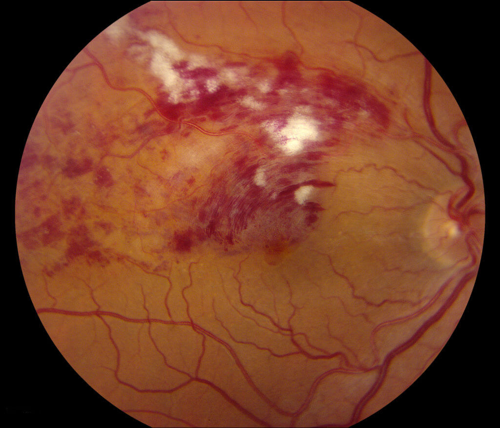
Most occlusions occur at an arteriovenous crossing or at the lamina cribrosa, where a stiff, sclerotic artery compresses the vein and slows flow. As a result, the retina shows the same arteriolosclerosis that affects cerebral vessels.
What the Evidence on RVO Stroke Risk Shows
A Korean nationwide cohort found more strokes after RVO than in matched controls, and a meta-analysis by Li and colleagues reported a similar association. Danish case-control data also linked branch occlusion with hypertension and cardiovascular disease. However, the excess risk appears modest, and shared risk factors explain much of it.
Fundus Explorer Pro
Photograph the retinal findings described here with the phone already in your pocket — 22 D optics and built-in illumination in one handheld unit.
From Choroida — the team behind this siteLimits of the Data
Most cohorts rely on insurance or registry coding, so misclassification is possible, and adjustment for blood pressure control and smoking also varies between studies. Therefore, avoid quoting a precise personal risk figure to the patient.
How RVO Differs From Artery Occlusion
Central retinal artery occlusion is an ocular stroke, often from carotid or cardiac emboli, so it needs same-day stroke evaluation (see retinal artery occlusion workup). Venous occlusion, by contrast, rarely requires emergency neurovascular imaging.
Risk Factors Behind RVO Stroke Risk
- Systemic hypertension, the strongest and most consistent association
- Diabetes mellitus
- Hyperlipidemia
- Smoking
- Obesity, and possibly obstructive sleep apnea
- Glaucoma or ocular hypertension
Baseline Tests in the RVO Stroke Risk Workup
First, measure blood pressure at the visit, and repeat it if the reading is high, then request HbA1c or fasting glucose and a lipid profile. A full blood count and renal function are also reasonable, so add ESR or plasma viscosity when the history suggests a hematologic or inflammatory cause.
Blood Pressure Comes First
Hypertension is the risk factor found most often in RVO case-control studies, and many patients first learn about it in the eye clinic. However, one raised reading does not establish the diagnosis, so ask primary care to confirm it with repeated or ambulatory measurements.
Who Needs Urgent Referral
Send patients with very high blood pressure plus headache, confusion or fellow eye hypertensive retinopathy the same day. New weakness, speech change or facial droop also needs emergency stroke services, and bilateral simultaneous occlusions suggest hyperviscosity and need prompt hematologic testing.
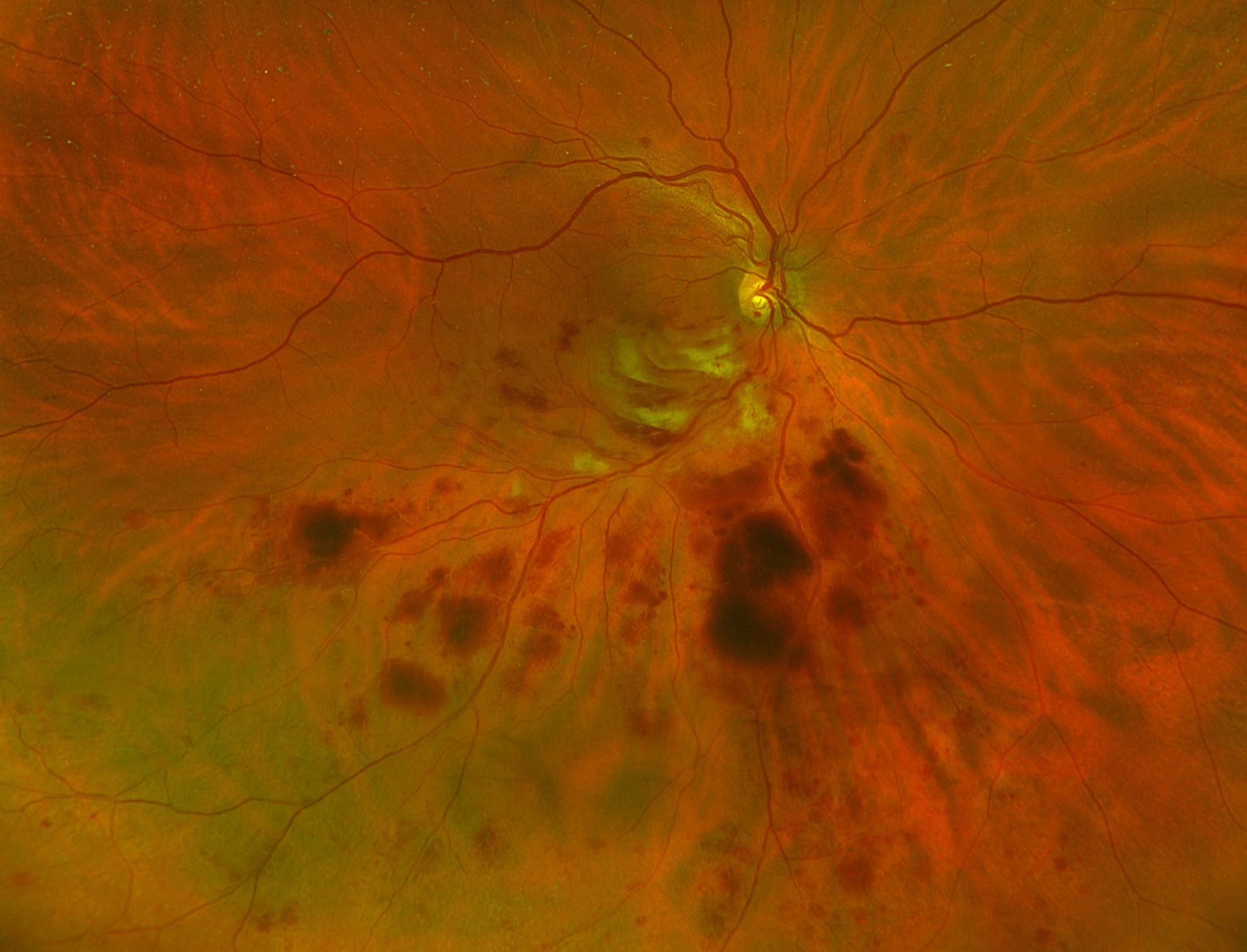
RVO Stroke Risk in Younger Patients
Patients under about 50 often lack classic vascular risk factors, so in this group the workup widens to thrombophilia, antiphospholipid antibodies and inflammatory disease (see RVO in young patients). Ask directly about oral contraceptives and dehydration as well, although routine thrombophilia panels in older patients rarely change management.
Carotid Imaging and RVO Stroke Risk
Carotid Doppler is not routine after a typical RVO, because venous occlusion does not arise from emboli. Nevertheless, order it for carotid symptoms, a bruit or features of ocular ischemic syndrome, and let mid-peripheral blot hemorrhages with dilated, nontortuous veins raise that suspicion.
Antiplatelet Therapy and RVO Stroke Risk
No convincing evidence shows that aspirin improves visual outcome after RVO, and some authors also suggest that antiplatelet drugs may increase retinal hemorrhage. Therefore, start aspirin or anticoagulation only for an established systemic indication, and the physician managing cardiovascular risk should make that decision.
Counseling the Patient on RVO Stroke Risk
Explain that the eye clot reflects vessel health and differs from a stroke. Then say plainly that later stroke and heart disease risk may be somewhat higher, though most of that risk responds to blood pressure, lipid and glucose control. Patients also act on concrete goals, such as stopping smoking and booking a primary care review.
Glaucoma and the Fellow Eye
The fellow eye can develop an occlusion over time, and glaucoma raises that risk, so check pressure and both optic discs (see glaucoma and retinal vein occlusion). Good systemic control may also protect the fellow eye, while direct proof remains limited.
Follow-Up Visits and RVO Stroke Risk
Send a short letter to primary care with the diagnosis, blood pressure and tests done. At each injection or monitoring visit, ask whether that review happened, and also ask about new neurological symptoms, chest pain or transient visual loss.
Pitfalls to Avoid
- Do not call a venous occlusion a stroke in the eye
- Never skip blood pressure measurement at diagnosis
- Avoid routine thrombophilia panels in older patients with clear vascular risk
- Do not start aspirin solely to treat the occlusion
- Remember that bilateral or atypical cases need a wider workup


Document what you see
Two smartphone imaging tools built for everyday clinic use — one for the slit lamp, one for the fundus.
From Choroida — the team behind this siteReferences
- Rim TH, Kim DW, Han JS, Chung EJ. Retinal vein occlusion and the risk of stroke development: a 9-year nationwide population-based study. Ophthalmology. 2015;122:1187-1194.
- Li M, Hu X, Huang J, Tan Y, Yang B, Tang Z. Impact of retinal vein occlusion on stroke incidence: a meta-analysis. J Am Heart Assoc. 2016;5:e004703.
- O’Mahoney PR, Wong DT, Ray JG. Retinal vein occlusion and traditional risk factors for atherosclerosis. Arch Ophthalmol. 2008;126:692-699.
- Bertelsen M, Linneberg A, Christoffersen N, et al. Comorbidity in patients with branch retinal vein occlusion: case-control study. BMJ. 2012;345:e7885.
- The Eye Disease Case-Control Study Group. Risk factors for central retinal vein occlusion. Arch Ophthalmol. 1996;114:545-554.
- Flaxel CJ, Adelman RA, Bailey ST, et al. Retinal Vein Occlusions Preferred Practice Pattern. Ophthalmology. 2020;127:P288-P320.

Writes and reviews the clinical articles on Educate. Every post is checked against peer-reviewed sources before it goes live.