Schirmer test results estimate aqueous tear production by measuring how far tears wet a filter paper strip.
Otto Schirmer described the method in 1903, and clinicians still use it daily.
However, the number only helps when you know which version the examiner used and why.
What the Schirmer Test Measures
The strip absorbs tears from the lower conjunctival sac over a fixed time.
Therefore, the wetted length reflects the volume of aqueous tear available.
The main lacrimal gland supplies most of this fluid, with help from accessory glands.
However, the test says little about lipid quality or tear film stability.
Schirmer I Without Anesthesia
Schirmer I uses no anesthetic drop.
As a result, it captures basal secretion plus reflex tearing that the strip itself provokes.
Most clinicians read it at 5 minutes with the eyes gently closed.
All-fit Slit-Lamp Adapter
Record and share exactly what you see at the slit lamp. One adapter fits any slit lamp or surgical microscope — and any smartphone.
From Choroida — the team behind this siteThe Anesthetized Basal Secretion Test
This variant aims to reduce the reflex component with topical anesthesia.
First, instill an anesthetic drop and wait about a minute.
Next, gently dry the inferior fornix so residual fluid does not inflate the reading.
Values run lower than Schirmer I, although reflex input rarely disappears completely.
Schirmer II and Reflex Tearing
Schirmer II also starts with topical anesthesia.
Then the examiner stimulates the nasal mucosa with a cotton applicator.
This checks whether the reflex arc can still drive lacrimal secretion.
A low result suggests severe gland failure, for example in advanced Sjögren disease.
How to Perform the Schirmer Test
- Perform it last, after osmolarity, break-up time and staining
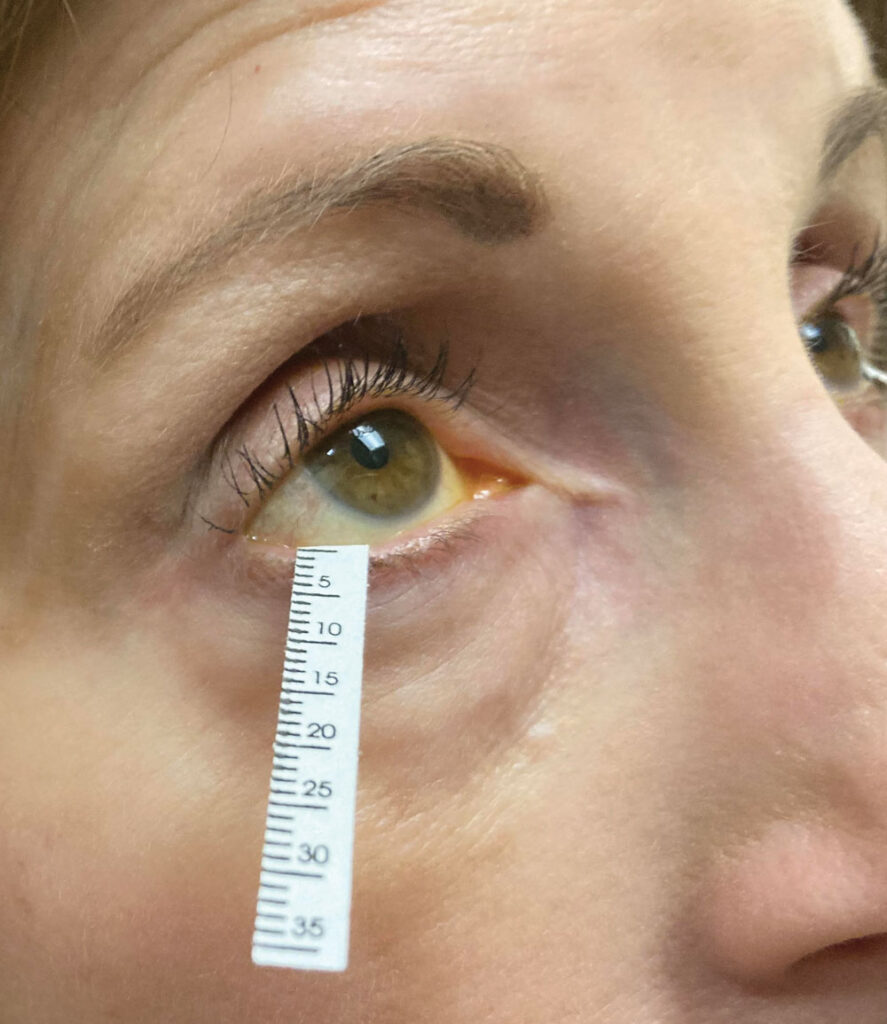
- Use standardized 5 x 35 mm strips folded at the notch
- Place the folded end at the junction of the middle and lateral thirds of the lower lid
- Avoid touching the cornea with the strip
- Remove strips at 5 minutes and read from the notch
Normal Schirmer Test Values
Without anesthesia, most clinicians accept 10 mm or more in 5 minutes as normal.
In contrast, values of 5 mm or less strongly suggest aqueous deficiency.
Results between 5 and 10 mm sit in a gray zone and need context.
With anesthesia, many examiners treat 5 mm or less as abnormal, although sources differ.
Schirmer Test Cut-Offs in Sjögren Disease
The 2016 ACR/EULAR criteria include a Schirmer result of 5 mm or less in 5 minutes in at least one eye.
This item scores one point, as does an ocular staining score of 5 or more.
However, these criteria classify patients for research and trials.
Therefore, they cannot replace clinical judgment in an individual patient.
Why Results Vary Between Visits
The Schirmer test shows limited repeatability from one visit to the next.
For example, Nichols and colleagues reported substantial variability in several common dry eye measurements, Schirmer included.
Reflex tearing also shifts with anxiety, light, humidity and lid position.
In addition, values often decline with age, so a single cut-off fits all ages poorly.
Sources of Error in the Schirmer Test
Residual anesthetic or earlier drops in the fornix can falsely raise the reading.
Strip contact with the cornea also triggers reflex tearing.
Similarly, squeezing, rapid blinking or a displaced strip distorts the result.
Finally, measure from the notch, because reading from the tip overstates wetting.
Schirmer Test Versus Tear Break-Up Time
Tear break-up time measures tear film stability (see tear break-up time).
A short break-up time with normal Schirmer wetting suggests evaporative dry eye.
Meibomian gland dysfunction is the usual driver of that pattern.
In contrast, low wetting points toward aqueous deficiency, while many patients show both.
Therefore, a normal Schirmer value never excludes dry eye.
Where Tear Osmolarity Fits
Tear osmolarity reflects the hyperosmolar state shared by both subtypes (see tear osmolarity testing).
DEWS II lists it among homeostasis markers, together with break-up time and ocular surface staining.
Schirmer does not appear in that core diagnostic battery.
Instead, the report uses tear volume measures to help subclassify dry eye after diagnosis.
Interpreting a Low Schirmer Test
A low reading should prompt a search for causes of aqueous deficiency.
Consider Sjögren disease, anticholinergic medications, prior refractive surgery and chronic lacrimal gland inflammation (see dacryoadenitis).
Then correlate it with staining, symptoms and systemic clues such as dry mouth.
Alternatives to the Schirmer Test
The phenol red thread test places a fine cotton thread in the lower fornix for 15 seconds.
Because it irritates less, it may provoke less reflex tearing.
Meanwhile, tear meniscus height on anterior segment OCT offers a noncontact estimate of tear volume.
However, neither method has displaced Schirmer in routine practice.
Pitfalls to Avoid
- Do not run Schirmer before break-up time or osmolarity
- Never compare anesthetized and unanesthetized values across visits
- Avoid diagnosing dry eye from one borderline reading
- Record the version, timing and eye position every time


Document what you see
Two smartphone imaging tools built for everyday clinic use — one for the slit lamp, one for the fundus.
From Choroida — the team behind this siteReferences
- Wolffsohn JS, Arita R, Chalmers R, et al. TFOS DEWS II diagnostic methodology report. Ocul Surf. 2017;15:539-574.
- Craig JP, Nichols KK, Akpek EK, et al. TFOS DEWS II definition and classification report. Ocul Surf. 2017;15:276-283.
- Shiboski CH, Shiboski SC, Seror R, et al. 2016 American College of Rheumatology/European League Against Rheumatism classification criteria for primary Sjögren’s syndrome. Arthritis Rheumatol. 2017;69:35-45.
- Nichols KK, Mitchell GL, Zadnik K. The repeatability of clinical measurements of dry eye. Cornea. 2004;23:272-285.
- American Academy of Ophthalmology. Basic and Clinical Science Course, Section 8: External Disease and Cornea. San Francisco: AAO; 2024.

Writes and reviews the clinical articles on Educate. Every post is checked against peer-reviewed sources before it goes live.