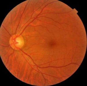
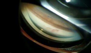
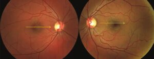
VKH vs sympathetic ophthalmia is a classic comparison because both cause bilateral granulomatous panuveitis.
Their eye findings overlap heavily, so the fundus alone rarely settles the question.
However, history, systemic features and timing usually separate them.
VKH vs Sympathetic Ophthalmia: The Core Difference
Sympathetic ophthalmia follows penetrating trauma or intraocular surgery to one eye.
In contrast, VKH develops without any such event.
Both diseases probably reflect T cell autoimmunity against melanocyte-associated antigens.
Taking the Trauma and Surgery History
Ask about every past injury, including childhood injuries and retained foreign bodies.
Next, ask about all eye operations, especially vitrectomy.
Patients often forget remote events, so look for scleral scars and iris defects.
Fundus Explorer Pro
Photograph the retinal findings described here with the phone already in your pocket — 22 D optics and built-in illumination in one handheld unit.
From Choroida — the team behind this siteWho Gets Each Disease
VKH is reported more often in pigmented populations, including Asian, Hispanic and Middle Eastern patients.
However, sympathetic ophthalmia shows no clear ethnic pattern, because it depends on the preceding injury.
Also, VKH typically presents in adults, so a young child with bilateral uveitis needs a broader differential.
Timing in VKH vs Sympathetic Ophthalmia
Sympathetic ophthalmia usually starts within months of the inciting event.
However, reported intervals range from days to decades, so a distant injury still counts.
VKH has no such anchor, because it moves through prodromal, acute uveitic, convalescent and chronic recurrent phases.
Neurologic and Auditory Clues
Headache, neck stiffness, tinnitus and dysacusis often precede visual loss in VKH.
Cerebrospinal fluid may show pleocytosis during this phase.
By comparison, such features are uncommon in sympathetic ophthalmia.
Skin and Hair Findings
Vitiligo, poliosis and alopecia support VKH when they accompany typical eye disease.
However, they usually appear during convalescence, so their absence at presentation does not exclude the diagnosis.
Slit Lamp Findings in VKH vs Sympathetic Ophthalmia
Both diseases can produce mutton-fat keratic precipitates, iris nodules and posterior synechiae.
However, early VKH may show mild anterior inflammation with marked posterior disease.
Ciliary body swelling can also shallow the chamber and induce myopia, which can mimic angle closure.
Examination Pearls
First, compare both eyes, because the fellow eye may show subtle flare before the patient notices symptoms.
Next, record intraocular pressure at every visit, since ciliary body edema and later synechiae can push it in either direction.
Also, dilate with care when the chamber is shallow.
Fundus Findings
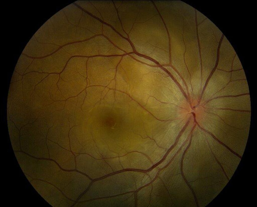
Multifocal exudative retinal detachments and disc hyperemia dominate acute VKH.
Sympathetic ophthalmia more often shows vitritis and yellow-white choroidal lesions.
Dalen-Fuchs nodules occur in both, so they do not discriminate.
Imaging in VKH vs Sympathetic Ophthalmia
Fluorescein angiography in acute VKH shows pinpoint leaks with later subretinal pooling.
Indocyanine green often shows hypofluorescent dark dots in both diseases (see modern VKH imaging).
Enhanced depth OCT shows a thickened choroid, and septated serous detachments strongly suggest acute VKH.
B-scan ultrasound helps when media are hazy, and it also helps exclude posterior scleritis.
Laboratory Testing
No blood test confirms either disease.
Instead, testing should exclude infection and other granulomatous causes (see uveitis testing).
Syphilis, tuberculosis and sarcoidosis deserve particular attention.
Other Diagnoses to Exclude
Several conditions can resemble either disease:
- Sarcoid uveitis, with bilateral granulomatous inflammation
- Syphilitic and tuberculous uveitis, which need antimicrobial treatment
- Posterior scleritis, with pain and exudative detachment
- Lens-induced uveitis, especially after trauma or surgery
Treatment in VKH vs Sympathetic Ophthalmia
Both diseases need prompt, high-dose systemic corticosteroids after infection has been excluded.
VKH generally requires a slow taper over at least six months, because early withdrawal invites recurrence.
Immunomodulatory therapy is often added for steroid sparing, but it needs laboratory monitoring.
These drugs cannot reverse established subretinal fibrosis or photoreceptor loss.
The Inciting Eye Question
Older retrospective data suggested a possible benefit from early enucleation, but the evidence is weak.
Therefore, consider removal mainly for a blind, painful eye with no visual potential.
Prognosis and Relapse
Early, adequate treatment often preserves useful vision in both diseases.
However, delayed or short courses raise the risk of chronic recurrence and a sunset glow fundus.
Also, sympathetic ophthalmia can relapse during tapering, so plan follow-up over several years.
Counseling and Follow-Up
Tell patients with injured or operated eyes to report fellow-eye redness, photophobia or blur immediately.
At follow-up, check for cataract, glaucoma and subretinal fibrosis.
See the existing reviews of VKH and sympathetic ophthalmia for more detail.
Pitfalls in VKH vs Sympathetic Ophthalmia
- Do not label bilateral panuveitis as VKH before asking about old surgery
- However, a trauma from decades earlier can still matter
- Also, absent skin changes at onset do not exclude VKH
- Never start high-dose steroids before considering syphilis and tuberculosis


Document what you see
Two smartphone imaging tools built for everyday clinic use — one for the slit lamp, one for the fundus.
From Choroida — the team behind this siteReferences
- Read RW, Holland GN, Rao NA, et al. Revised diagnostic criteria for Vogt-Koyanagi-Harada disease: report of an international committee on nomenclature. Am J Ophthalmol. 2001;131:647-652.
- Moorthy RS, Inomata H, Rao NA. Vogt-Koyanagi-Harada syndrome. Surv Ophthalmol. 1995;39:265-292.
- Kilmartin DJ, Dick AD, Forrester JV. Prospective surveillance of sympathetic ophthalmia in the UK and Republic of Ireland. Br J Ophthalmol. 2000;84:259-263.
- Lubin JR, Albert DM, Weinstein M. Sixty-five years of sympathetic ophthalmia. Ophthalmology. 1980;87:109-121.
- Chang GC, Young LH. Sympathetic ophthalmia. Semin Ophthalmol. 2011;26:316-320.

Writes and reviews the clinical articles on Educate. Every post is checked against peer-reviewed sources before it goes live.