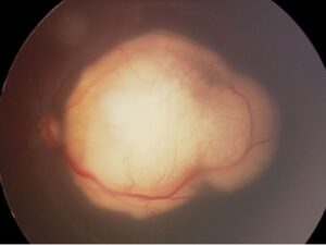
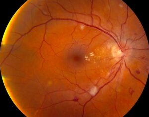
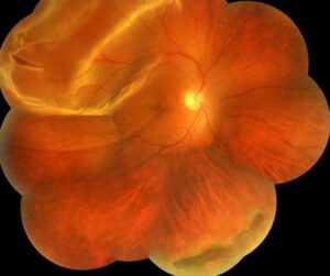
Drusen vs exudates is a common diagnostic question when yellow spots appear in the macula.
Drusen lie beneath the retinal pigment epithelium (RPE), while hard exudates lie within the neurosensory retina.
Therefore, the two lesions point toward different diseases and different management.
Drusen vs Exudates at a Glance
Four features usually settle the question: color, pattern, OCT level and associated disease.
First, drusen tend to look dull and deep, whereas exudates look bright and waxy.
Next, OCT places each lesion in a different layer.
Finally, the clinical context usually confirms the impression.
What Drusen Are
Drusen are focal extracellular deposits containing lipid, protein and cellular debris.
They accumulate between the RPE basement membrane and Bruch’s membrane.
Small hard drusen often occur with normal aging, while large soft drusen carry greater risk (see soft and hard drusen).
Also, clinical classification uses drusen size to grade age-related macular degeneration (AMD).
Fundus Explorer Pro
Photograph the retinal findings described here with the phone already in your pocket — 22 D optics and built-in illumination in one handheld unit.
From Choroida — the team behind this siteWhat Hard Exudates Are
Hard exudates form when damaged vessels leak lipoproteins into the retina.
Because fluid reabsorbs faster than lipid, yellow deposits remain behind.
These deposits usually collect in the outer plexiform layer.
Therefore, each exudate suggests a current or recent leak nearby (see hard exudates).
Drusen vs Exudates on Fundus Examination
Drusen appear pale yellow, with discrete or soft borders.
They look deep to the retinal vessels on stereoscopic examination.
In contrast, exudates often glisten and have sharp, irregular margins.
Surrounding retinal thickening also favors exudates, particularly when microaneurysms or hemorrhages lie nearby.
Pattern and Distribution
Drusen often scatter across both maculae in a roughly symmetric pattern.
Exudates follow the leak, so they may form a circinate ring around leaking microaneurysms.
For example, a macroaneurysm can produce a dense ring or plaque in one eye.
A macular star suggests lipid deposition along Henle fibers, as in neuroretinitis or severe hypertension.
Drusen vs Exudates on OCT
OCT usually gives the clearest answer.
Drusen elevate the RPE and create dome-shaped or undulating bumps above Bruch’s membrane.
However, exudates appear as hyperreflective foci or plaques within the outer retina, often with posterior shadowing.
Intraretinal cysts and retinal thickening may accompany them, which supports active leakage.
Diseases Associated With Drusen
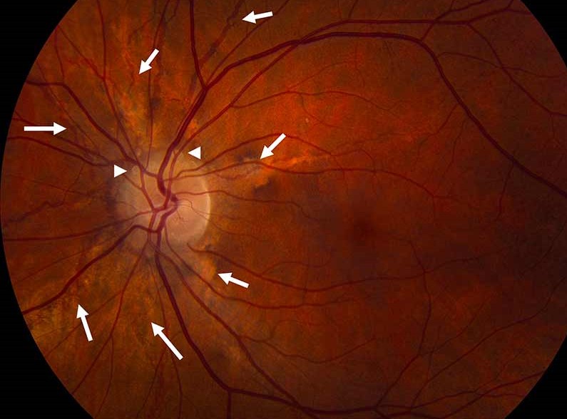
AMD is the main association in patients over 50 years of age.
However, younger patients with numerous small drusen may have cuticular drusen or an inherited macular disease.
Family history and multimodal imaging then become more useful.
Smoking history also matters, because it raises AMD progression risk.
Diseases Associated With Exudates
Diabetic retinopathy is the most frequent cause in routine practice.
Other causes include retinal vein occlusion, macroaneurysm, Coats disease, radiation retinopathy and hypertensive retinopathy.
In addition, higher serum lipid levels were associated with more hard exudate in the ETDRS cohort.
Therefore, ask about glucose, blood pressure and lipid control.
When Both Lesions Coexist
An older patient with drusen and new exudates needs prompt evaluation for macular neovascularization.
OCT may reveal subretinal fluid or a pigment epithelial detachment, while OCT angiography can show the abnormal network.
A patient with diabetes can also develop AMD.
As a result, each lesion deserves its own explanation.
Other Yellow or White Lookalikes
Several other lesions can mimic either finding.
For example, cotton wool spots look white and fluffy, and they lie in the nerve fiber layer.
Stargardt flecks, pattern dystrophy deposits and crystalline maculopathy also enter the differential.
Then, autofluorescence and OCT help narrow the options.
Drusen vs Exudates and Treatment Decisions
No proven treatment safely removes drusen to prevent AMD progression.
AREDS2-based supplements may reduce progression in suitable patients, but they do not restore lost vision or cure AMD.
Exudates require treatment of the leak and its systemic drivers.
Options include anti-VEGF injections, intravitreal steroids or focal laser, each with its own risks.
Drusen vs Exudates at Follow-Up
Drusen follow-up looks for new fluid, hemorrhage or atrophy on OCT and examination.
Next, teach patients to report new distortion or central blur promptly.
For exudates, track retinal thickness, foveal involvement and the response to treatment.
Lipid may clear slowly over months, even after the leak improves.
Pitfalls to Avoid
- Do not call every yellow macular dot drusen without checking OCT
- Do not overlook neovascularization when exudates appear in an eye with AMD
- Avoid treating exudates without searching for the leaking source
- Do not assume bilateral lesions are always drusen, because diabetes often affects both eyes


Document what you see
Two smartphone imaging tools built for everyday clinic use — one for the slit lamp, one for the fundus.
From Choroida — the team behind this siteReferences
- Curcio CA, Millican CL. Basal linear deposit and large drusen are specific for early age-related maculopathy. Arch Ophthalmol. 1999;117:329-339.
- Ferris FL 3rd, Wilkinson CP, Bird A, et al. Clinical classification of age-related macular degeneration. Ophthalmology. 2013;120:844-851.
- Bolz M, Schmidt-Erfurth U, Deak G, et al. Optical coherence tomography hyperreflective foci: a morphologic sign of lipid extravasation in diabetic macular edema. Ophthalmology. 2009;116:914-920.
- Chew EY, Klein ML, Ferris FL 3rd, et al. Association of elevated serum lipid levels with retinal hard exudate in diabetic retinopathy: ETDRS report 22. Arch Ophthalmol. 1996;114:1079-1084.
- Age-Related Eye Disease Study 2 Research Group. Lutein + zeaxanthin and omega-3 fatty acids for age-related macular degeneration. JAMA. 2013;309:2005-2015.

Writes and reviews the clinical articles on Educate. Every post is checked against peer-reviewed sources before it goes live.