
Pseudomonas keratitis is a bacterial corneal infection caused by Pseudomonas aeruginosa, and it is the most aggressive of the common bacterial ulcers.
It is closely linked to contact lens wear, particularly overnight wear, and it can destroy a cornea within days if it is not treated.
Early recognition and intensive therapy are what separate a healed scar from a perforation.
What Is Pseudomonas Keratitis?
P. aeruginosa is a gram-negative rod that thrives in moist environments, including lens cases, solutions, and water.
It adheres to compromised epithelium and releases proteases and other enzymes that dissolve the stroma.
Contact lens wear is the leading risk factor, and the others include trauma, ocular surface disease, previous surgery, and hospitalization with intubation or an ocular exposure (see contact lens-related microbial keratitis).
The same organism is a frequent cause of keratitis in contaminated ophthalmic solutions and in intensive care patients.
Clinical Features
The onset is sudden, with pain, redness, photophobia, tearing, and rapidly reduced vision.
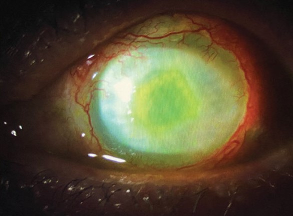
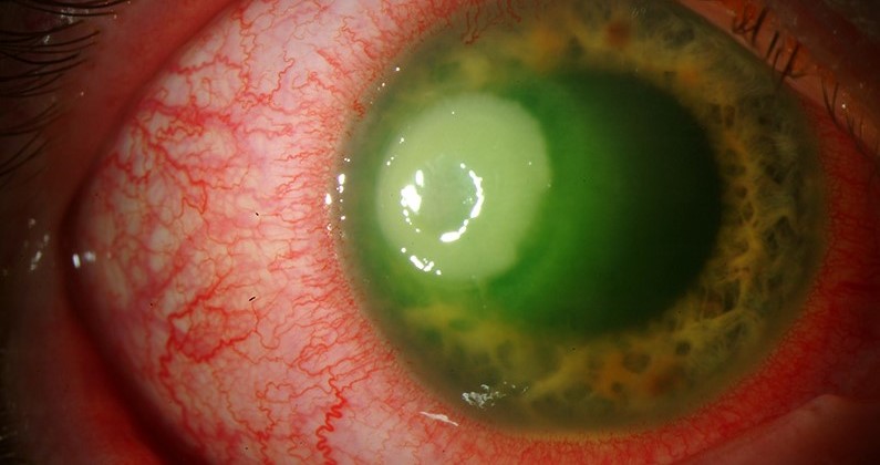
The ulcer is typically central or paracentral, with a gray-white infiltrate that expands over hours.
A surrounding ring of edema and a mucopurulent, greenish discharge are characteristic, and they result from the pigment and exudate of the organism.
A hypopyon forms early, and the stroma looks “ground-glass” and necrotic.
Without treatment, the cornea can thin and perforate within a few days.
All-fit Slit-Lamp Adapter
Record and share exactly what you see at the slit lamp. One adapter fits any slit lamp or surgical microscope — and any smartphone.
From Choroida — the team behind this siteDiagnosis
Take a corneal scraping for Gram stain and culture before starting treatment when it is practical to do so.
The Gram stain shows gram-negative rods, and culture on blood and chocolate agar produces typical colonies with a metallic sheen and a sweet odor.
A culture should also be taken from the contact lens and the lens case if the patient has them.
Treatment should not wait for culture results in a clinically typical case.
Differential Diagnosis
- Other bacterial ulcers: Staphylococcus and Streptococcus produce a more discrete, less rapidly expanding infiltrate
- Fungal keratitis: a slower course, feathery margins, and a history of plant trauma
- Acanthamoeba keratitis: severe pain with a ring infiltrate in a lens wearer (see Acanthamoeba keratitis)
- Sterile infiltrates: smaller, peripheral, with minimal pain and no epithelial defect
Treatment
Treatment starts the same day with intensive topical antibiotics, usually a fluoroquinolone, or fortified tobramycin or gentamicin combined with a cephalosporin in a severe case.
Dosing is hourly, including at night for the first period, and the response is judged after one to two days.
Cycloplegics reduce pain and prevent synechiae, and a systemic antibiotic or a collagenase inhibitor may be added when the cornea is melting (see corneal melt).
The role of steroids is discussed after the organism is controlled, and the evidence for benefit is limited.
Perforation or impending perforation calls for tissue adhesive, a patch graft, or keratoplasty.
Patients should be told to stop wearing lenses and to discard the case, and to seek care promptly if symptoms recur.
The Role of Contact Lens Hygiene
Overnight wear of lenses increases the risk of microbial keratitis several-fold, and Pseudomonas is the organism most often associated with it.
Lens cases are a common reservoir, and a biofilm may form on the lens surface, which protects the bacteria from disinfectants.
Rinsing lenses with tap water, topping up old solution, and failing to replace the case at regular intervals are common habits that bring bacteria to the eye.
At the visit, ask the patient about their habits specifically, and also whether they clean their lenses.
What the Cornea Looks Like Over Time
In the first day the infiltrate is small and the epithelium is the main focus, and by the second or third day the stroma may be liquefied and edematous.
A central ulcer with a ring-shaped infiltrate, expanding rapidly, is a sign of a severe infection.
Descemetocele and perforation are late events that are avoided by early, intensive treatment.
A residual central scar is common, and some patients later need a rigid contact lens or keratoplasty for visual rehabilitation.
Monitoring Response
Review the patient within 24 hours of starting therapy and measure the infiltrate with the slit-lamp scale.
An ulcer that has stopped enlarging after two days is usually responding, even if the appearance has not yet improved.
Persistent enlargement, a rising hypopyon, or thinning calls for a repeat culture, a change of regimen, and consideration of corneal cross-linking or surgical options in specialist centers.
A written plan with the dosing schedule, the next appointment, and a number to call helps the patient to follow the intensive regimen.
Admission to hospital is reasonable when the ulcer is large, when the patient cannot manage hourly drops, or when adherence is a concern.


Document what you see
Two smartphone imaging tools built for everyday clinic use — one for the slit lamp, one for the fundus.
From Choroida — the team behind this siteReferences
- Hazlett LD. Corneal response to Pseudomonas aeruginosa infection. Prog Retin Eye Res. 2004;23:1-30.
- Green M, Apel A, Stapleton F. Risk factors and causative organisms in microbial keratitis. Cornea. 2008;27:22-27.
- Mannis MJ, Holland EJ, eds. Cornea. 4th ed. Elsevier; 2017.
- American Academy of Ophthalmology. Basic and Clinical Science Course, Section 8: External Disease and Cornea. AAO; current edition.

Writes and reviews the clinical articles on Educate. Every post is checked against peer-reviewed sources before it goes live.


