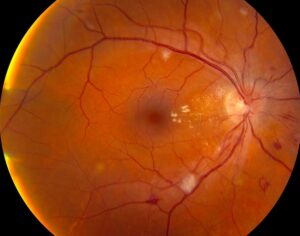
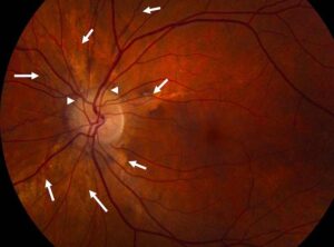
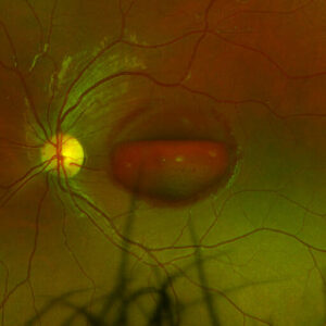
Retinal detachment recovery usually takes months, even when the retina reattaches after one operation.
Anatomic success comes first, while visual function improves more slowly.
Therefore, counseling should separate these two goals from the first postoperative visit.
The Retinal Detachment Recovery Timeline
The first week centers on comfort, positioning and pressure control.
Then, over several weeks, inflammation settles and any gas bubble shrinks.
Vision often keeps improving for months, and some eyes continue gaining beyond the first year.
However, timelines vary widely, so avoid promising a fixed date.
The First Postoperative Days
Patients commonly report redness, lid swelling, tearing and a gritty sensation.
Mild aching usually responds to oral analgesics.
Severe pain, however, suggests raised pressure or infection and needs same-day review.
Fundus Explorer Pro
Photograph the retinal findings described here with the phone already in your pocket — 22 D optics and built-in illumination in one handheld unit.
From Choroida — the team behind this siteMacular Status and Retinal Detachment Recovery
Preoperative macular status strongly influences the final result.
Macula-on eyes often retain good acuity, although floaters and mild distortion may persist.
In contrast, macula-off eyes recover more slowly, and many never regain their previous vision (see macula-off recovery).
OCT helps explain this variation because outer retinal band disruption correlates with poorer acuity after repair.
Positioning During Retinal Detachment Recovery
Positioning aims to hold the tamponade against the break while the retinopexy adhesion matures.
Surgeons therefore tailor posture to break location, tamponade type and macular status.
Patients also need written instructions that state the posture, daily hours and total duration.
Vague advice tends to produce poor adherence, especially in older patients with neck or back disease.
What Positioning Trials Show
The PostRD randomized trial studied macula-involving detachments repaired with vitrectomy and gas.
Face-down positioning reduced retinal displacement compared with support-the-break positioning.
However, protocols still differ among surgeons, and the ideal duration remains uncertain.
Patients should therefore follow their own surgeon’s plan, even when a friend received different advice.
Gas Bubble Limits in Retinal Detachment Recovery
Intraocular gas expands when ambient pressure falls.
As a result, patients must avoid flying and high-altitude travel until the surgeon confirms the bubble has gone.
Nitrous oxide anesthesia also expands the bubble and can raise pressure enough to occlude the central retinal artery.
Therefore, patients should wear a gas warning wristband and tell every anesthesia provider.
How Long the Gas Lasts
Duration depends on the gas and its concentration.
SF6 typically lasts about two weeks, while C3F8 can last about two months.
At first, vision looks very blurred through the bubble.
Later, patients see a moving line that sinks lower as the gas absorbs.
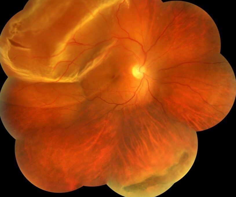
Silicone Oil and Retinal Detachment Recovery
Silicone oil gives longer tamponade, so vision often stays limited while it remains.
Unlike gas, oil does not carry the same altitude restriction.
However, it can cause pressure rise, emulsification, cataract and corneal problems.
Removal timing is individualized, and redetachment can still occur after removal.
Recovery After Scleral Buckle
A buckle avoids intraocular tamponade in many cases, so some patients skip gas restrictions.
However, the encircling element can induce a myopic shift.
Early pain and lid swelling may be more noticeable, and some patients develop diplopia.
PIVOT also showed better 12-month acuity with pneumatic retinopexy than vitrectomy in selected eyes, so method can shape recovery.
Complications That Slow Retinal Detachment Recovery
Cataract commonly progresses after vitrectomy in phakic eyes, and later surgery often restores useful vision.
Epiretinal membrane can also cause progressive distortion.
In addition, cystoid changes may limit acuity (see macular edema after repair).
OCT separates these causes, so obtain it whenever vision stalls or declines.
Recurrence and Follow-Up
Proliferative vitreoretinopathy is a leading cause of late failure, and it typically appears within the first months.
Therefore, careful peripheral examination continues after the retina looks attached.
New breaks can also cause recurrence, sometimes after apparently stable healing (see recurrent detachment).
Next, remember the fellow eye, which deserves dilated examination and clear symptom advice.
Warning Signs After Surgery
Teach these symptoms at discharge and ask for urgent contact if any appear:
- Increasing pain, especially with nausea or headache
- Falling vision instead of gradual improvement
- A new curtain or shadow in any direction
- New floaters or flashes
- Increasing redness, discharge or light sensitivity
Pitfalls to Avoid
- Do not equate an attached retina with normal vision
- Never clear a patient for flying without confirming that the gas has gone
- Do not attribute falling vision to the bubble without examining the eye


Document what you see
Two smartphone imaging tools built for everyday clinic use — one for the slit lamp, one for the fundus.
From Choroida — the team behind this siteReferences
- Heimann H, Bartz-Schmidt KU, Bornfeld N, et al. Scleral buckling versus primary vitrectomy in rhegmatogenous retinal detachment. Ophthalmology. 2007;114:2142-2154.
- Hillier RJ, Felfeli T, Berger AR, et al. The Pneumatic Retinopexy versus Vitrectomy for the Management of Primary Rhegmatogenous Retinal Detachment Outcomes Randomized Trial (PIVOT). Ophthalmology. 2019;126:531-539.
- Casswell EJ, Yorston D, Lee E, et al. Effect of face-down positioning vs support-the-break positioning after macula-involving retinal detachment repair: the PostRD randomized clinical trial. JAMA Ophthalmol. 2020;138:634-642.
- Shiragami C, Shiraga F, Yamaji H, et al. Unintentional displacement of the retina after standard vitrectomy for rhegmatogenous retinal detachment. Ophthalmology. 2010;117:86-92.
- Wakabayashi T, Oshima Y, Fujimoto H, et al. Foveal microstructure and visual acuity after retinal detachment repair. Ophthalmology. 2009;116:519-528.

Writes and reviews the clinical articles on Educate. Every post is checked against peer-reviewed sources before it goes live.