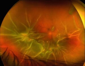
Salmon patch hemorrhage is an oval intraretinal bleed in the midperipheral fundus of patients with sickle cell disease.
It follows sudden occlusion of a small retinal arteriole.
The lesion changes color and shape over days to weeks, so one eye may show several stages at once.
Therefore, recognizing this evolution helps the examiner read the whole sickle fundus.
What Is a Salmon Patch Hemorrhage?
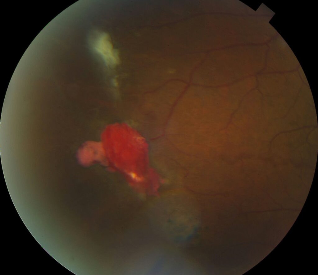
The lesion is a well-circumscribed hemorrhage lying next to a peripheral arteriole.
When fresh, it looks bright red.
Then, as hemoglobin breaks down, it turns orange-pink, which gives the sign its name.
Many patches resolve completely and leave a normal-looking retina.
Pathogenesis of Salmon Patch Hemorrhage
Sickled erythrocytes obstruct a medium-sized arteriole in the midperiphery.
As a result, the ischemic vessel wall weakens and ruptures.
Blood then dissects into the retina, and it may also break through the internal limiting membrane or extend beneath the retina.
Fundus Explorer Pro
Photograph the retinal findings described here with the phone already in your pocket — 22 D optics and built-in illumination in one handheld unit.
From Choroida — the team behind this siteWho Develops It
Salmon patches occur in hemoglobin SS and SC disease, and less often in other sickling genotypes.
Most patients have no visual symptoms because the lesions sit outside the macula.
Therefore, the sign usually appears as an incidental finding during dilated screening.
However, it confirms active vaso-occlusion in that eye.
Clinical Appearance of Salmon Patch Hemorrhage
Indirect ophthalmoscopy with scleral depression gives the best view.
Look for a flat or slightly dome-shaped hemorrhage beside an arteriole, often with a smooth outline.
In addition, the overlying retina may appear mildly elevated.
A preretinal component can form a boat-shaped layer, while a subretinal component looks darker.
Iridescent Spots After Salmon Patch Hemorrhage
Some lesions leave small glistening, refractile deposits on the inner retinal surface.
These iridescent spots contain hemosiderin-laden macrophages.
They often sit inside a shallow depression or cavity.
Because they persist for long periods, they act as a record of earlier bleeding.
Schisis Cavities in Salmon Patch Hemorrhage
As blood resorbs within the inner retina, it can leave a localized splitting of the layers.
Clinically, this looks like a small, sharply edged schisis cavity, often lined by iridescent spots.
Where the scan can reach the periphery, OCT may show inner retinal thinning or splitting.
These cavities usually stay stable and need observation only.
Black Sunburst and the Subretinal Route
Sometimes blood dissects beneath the neurosensory retina and stimulates the retinal pigment epithelium.
As a result, pigment epithelial hyperplasia and migration produce a black sunburst lesion.
These are round, pigmented peripheral patches with spiculated borders.
Salmon patches, iridescent spots and black sunbursts form the classic nonproliferative signs.
Salmon Patch Hemorrhage and Goldberg Staging
Goldberg staging describes proliferative disease, from peripheral arteriolar occlusion to retinal detachment (see Goldberg classification).
Salmon patches are nonproliferative, so they do not define a stage.
However, they arise from the same peripheral arteriolar closure that marks stage I.
Therefore, a patch should prompt a careful search for arteriovenous anastomoses and sea fans.
Imaging the Lesion
Widefield color photography records size and color for later comparison.
On fluorescein angiography, the blood causes blocked fluorescence.
In addition, angiography reveals peripheral nonperfusion and any neovascularization near the occluded arteriole.
Differential Diagnosis
- Valsalva or traumatic preretinal bleeding (see Valsalva retinopathy)
- Hemorrhages from severe anemia or leukemia
- Diabetic and retinal vein occlusion hemorrhages
- Roth spots in endocarditis
- Pigmented chorioretinal scars, which can mimic a black sunburst
Management of Salmon Patch Hemorrhage
The hemorrhage itself needs no treatment.
It resolves spontaneously, and laser offers no benefit for the patch.
Instead, management focuses on surveillance for proliferative change.
Then, treat sea fans and their complications according to the overall sickle cell retinopathy plan.
Follow-up and Monitoring
Repeat dilated examination with scleral depression at intervals that suit the genotype and findings.
Patients with HbSC disease need close review because proliferative disease is more common in that group.
Also, compare photographs to separate new hemorrhages from old ones.
Finally, a rising number of fresh patches suggests ongoing vaso-occlusion.
Counseling About Salmon Patch Hemorrhage
Explain that the lesion is a small bleed in the far edge of the retina.
It usually clears without treatment and rarely affects reading vision.
However, it shows that sickling already affects the retinal vessels.
Therefore, ask the patient to keep regular eye visits and to report floaters, a curtain or sudden blur at once.
Pitfalls to Avoid
- Do not laser a salmon patch, because it resolves on its own
- Never skip scleral depression, since sea fans hide in the far periphery
- Avoid mistaking iridescent spots for retinal crystals of another cause
- Do not assume a normal macula means a normal sickle fundus


Document what you see
Two smartphone imaging tools built for everyday clinic use — one for the slit lamp, one for the fundus.
From Choroida — the team behind this siteReferences
- Goldberg MF. Classification and pathogenesis of proliferative sickle retinopathy. Am J Ophthalmol. 1971;71:649-665.
- Gagliano DA, Goldberg MF. The evolution of salmon-patch hemorrhages in sickle cell retinopathy. Arch Ophthalmol. 1989;107:1814-1815.
- Romayanada N, Goldberg MF, Green WR. Histopathology of sickle cell retinopathy. Trans Am Acad Ophthalmol Otolaryngol. 1973;77:OP652-OP676.
- Elagouz M, Jyothi S, Gupta B, Sivaprasad S. Sickle cell disease and the eye: old and new concepts. Surv Ophthalmol. 2010;55:359-377.
- American Academy of Ophthalmology. Basic and Clinical Science Course, Section 12: Retina and Vitreous. San Francisco: AAO; latest edition.

Writes and reviews the clinical articles on Educate. Every post is checked against peer-reviewed sources before it goes live.