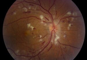
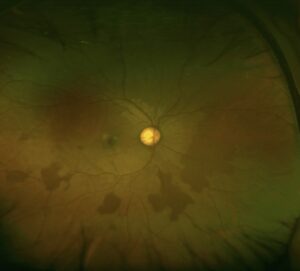
The Beckman classification stages age-related macular degeneration (AMD) in people older than 55 years.
It uses drusen size and pigment changes within two disc diameters of the fovea.
Because it needs only a dilated examination or color photograph, it suits routine practice.
What the Beckman Classification Measures
An expert committee published the scale in 2013.
Two findings drive staging: the largest druse and any associated pigment change.
Late disease then adds neovascular AMD and geographic atrophy.
Measuring Drusen at the Slit Lamp
The scale uses two thresholds, 63 and 125 microns.
For example, 125 microns roughly matches the width of a large vein at the disc margin.
A druse about half that width approaches 63 microns.
However, magnification varies, so compare each druse with that vein.
Morphology adds information (see soft and hard drusen).
Fundus Explorer Pro
Photograph the retinal findings described here with the phone already in your pocket — 22 D optics and built-in illumination in one handheld unit.
From Choroida — the team behind this siteThe Five Beckman Classification Stages
- No aging changes: no drusen and no pigmentary abnormalities
- Normal aging changes: only drupelets, smaller than 63 microns
- Early AMD: medium drusen (63 to 125 microns) without pigment abnormalities
- Intermediate AMD: large drusen, or pigment abnormalities with medium drusen
- Late AMD: neovascular AMD or any geographic atrophy
Normal Aging and Early AMD
Drupelets are common in older eyes and carry no clinically relevant risk.
Therefore, avoid giving these patients an AMD diagnosis.
Medium drusen define early AMD, and patients usually keep good acuity.
Nevertheless, discuss smoking, diet and periodic examination.
Intermediate AMD in the Beckman Classification
One large druse within the macular zone qualifies.
Pigment changes with at least medium drusen also qualify.
Some patients notice slow dark adaptation despite good acuity.
However, risk varies widely, because bilateral findings carry more weight.
Late AMD: Neovascular Disease and Atrophy
Late AMD includes neovascular disease or geographic atrophy of any size.
Examiners may see subretinal fluid, hemorrhage or a sharply demarcated atrophic patch.
OCT then confirms fluid, neovascular tissue and outer retinal loss.
Foveal involvement is separate, because atrophy can spare the center.
Progression Risk in the Beckman Classification
Five-year risk of late AMD rises about 100-fold across the scale.
It ranges from about 0.5% with normal aging changes to about 50% in the highest-risk group.
These figures describe groups, so they cannot predict one patient’s course.
Comparison With the AREDS Simplified Scale
This scale gives one point per eye for large drusen and one for pigment abnormalities.
Thus, it refines risk within the broad intermediate stage.
Five-year risks for scores 0 to 4 are about 0.5%, 3%, 12%, 25% and 50%.
Supplements and Beckman Classification Stage
AREDS reduced progression in participants with intermediate disease or late AMD in one eye.
Therefore, AREDS-type supplements suit those patients, while early disease lacks comparable evidence.
AREDS2 then supported lutein and zeaxanthin in place of beta-carotene.
Finally, explain that supplements slow risk modestly and do not reverse damage.
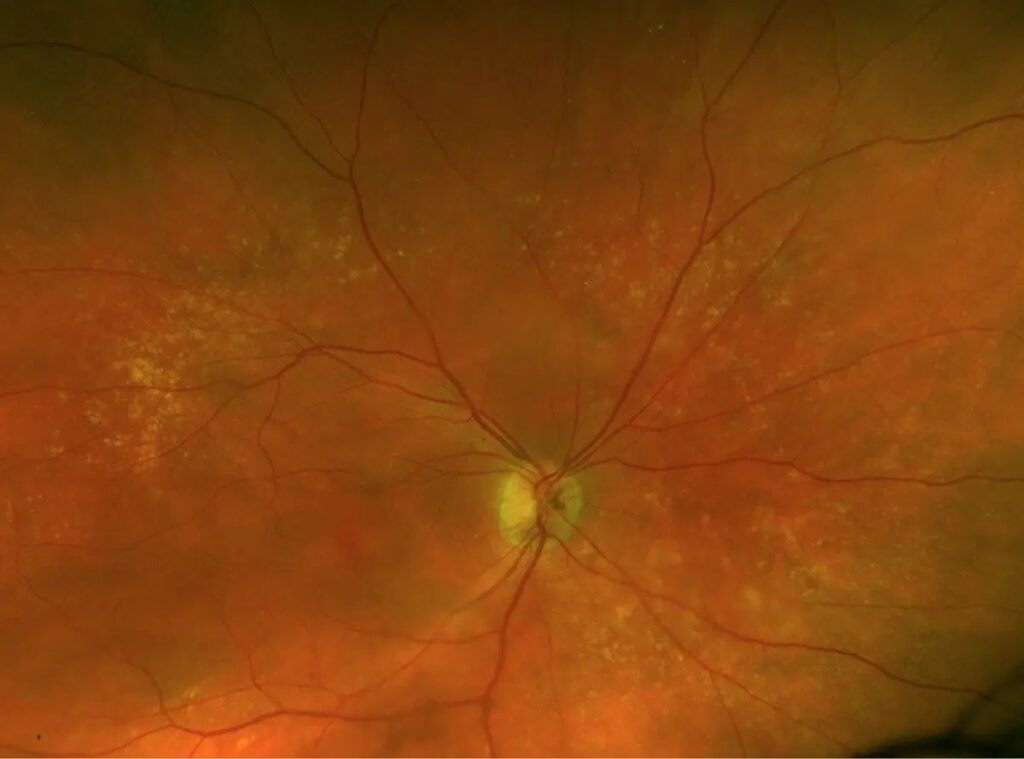
Management of Late Disease
Active neovascular AMD needs prompt anti-VEGF evaluation (see wet vs dry AMD).
Complement inhibitors may slow geographic atrophy growth in selected patients.
However, they do not restore lost vision.
Imaging Limits of the Beckman Classification
The scale predates modern multimodal grading.
Consequently, it does not stage reticular pseudodrusen, which OCT often reveals.
Consensus OCT definitions of atrophy now add structural detail.
Mimics That Distort Staging
For example, basal laminar drusen can appear as numerous small lesions in younger patients.
Pattern dystrophy and adult vitelliform lesions can also mislead.
Therefore, check age, family history, autofluorescence and OCT when the appearance is atypical.
Counseling Patients Using the Beckman Classification
Use plain language first, because many patients equate AMD with blindness.
AMD rarely causes complete blindness, although central vision can decline.
Next, name the stage and describe the approximate risk.
Also teach patients to report new distortion or sudden blur promptly.
At each visit, compare drusen, pigment and OCT with baseline.
Follow-Up After Staging
Examination intervals depend on stage, symptoms and fellow-eye status.
Patients with intermediate disease need clear self-monitoring instructions.
An Amsler grid may help, although a normal grid cannot exclude neovascularization.
Documenting the Beckman Classification Stage
Record the stage separately for each eye, because the fellow eye changes risk.
Also note the largest druse, pigment changes and the imaging used.
Then, compare the next visit with the same baseline photograph or OCT scan.
As a result, small changes become easier to recognize.
Risk Factors That Modify Staging Decisions
- Smoking, which raises progression risk and is modifiable
- Family history of AMD, because genetic risk is substantial
- Older age, since progression risk rises over time
- Fellow-eye late AMD, which increases risk in the second eye
Pitfalls to Avoid
- Do not label drupelets as early AMD
- Do not stage lesions outside the two-disc-diameter zone
- Avoid estimating drusen size without a reference vessel
- Do not assume geographic atrophy always involves the fovea
- Never recommend supplements without checking the stage


Document what you see
Two smartphone imaging tools built for everyday clinic use — one for the slit lamp, one for the fundus.
From Choroida — the team behind this siteReferences
- Ferris FL 3rd, Wilkinson CP, Bird A, et al. Clinical classification of age-related macular degeneration. Ophthalmology. 2013;120:844-851.
- Ferris FL, Davis MD, Clemons TE, et al. A simplified severity scale for age-related macular degeneration: AREDS report no. 18. Arch Ophthalmol. 2005;123:1570-1574.
- Age-Related Eye Disease Study 2 Research Group. Lutein + zeaxanthin and omega-3 fatty acids for age-related macular degeneration. JAMA. 2013;309:2005-2015.
- Sadda SR, Guymer R, Holz FG, et al. Consensus definition for atrophy associated with age-related macular degeneration on OCT. Ophthalmology. 2018;125:537-548.
Test yourself
A few questions straight from this article.
-
Within what zone are lesions graded in the Beckman classification of AMD?
The Beckman scale grades drusen size and pigment changes within two disc diameters of the fovea, and lesions outside that zone should not be staged. -
In the Beckman classification, which finding defines early AMD?
Early AMD is medium drusen of 63 to 125 microns without pigment abnormalities, and these patients usually keep good acuity. -
When staging AMD at the slit lamp, which landmark approximates the 125-micron drusen threshold?
125 microns roughly matches the width of a large vein at the disc margin, and a druse about half that width approaches 63 microns. -
Under the Beckman classification, an eye with only drupelets smaller than 63 microns is staged as what?
Drupelets alone are normal aging changes with no clinically relevant risk, so these patients should not be given an AMD diagnosis. -
In the Beckman classification, which single finding is enough for intermediate AMD?
One large druse within the macular zone qualifies as intermediate AMD, as do pigment changes with at least medium drusen. -
Under the Beckman classification, how much geographic atrophy is needed to classify an eye as late AMD?
Late AMD includes neovascular AMD or geographic atrophy of any size; foveal involvement is recorded separately because atrophy can spare the center. -
Across the Beckman AMD scale, roughly how much does the five-year risk of late AMD increase?
Five-year risk rises about 100-fold across the scale, from about 0.5% with normal aging changes to about 50% in the highest-risk group. -
Which findings earn points on the AREDS simplified severity scale for AMD?
The simplified scale gives one point per eye for large drusen and one for pigment abnormalities, refining risk within the intermediate stage. -
Based on AREDS, which patients with AMD are suited to AREDS-type supplements?
AREDS reduced progression in intermediate disease or late AMD in one eye, while early disease lacks comparable evidence, so the stage should be checked first. -
In AREDS2, which nutrients were supported as a replacement for beta-carotene in AMD supplements?
AREDS2 supported lutein and zeaxanthin in place of beta-carotene; supplements slow risk modestly and do not reverse damage.

Writes and reviews the clinical articles on Educate. Every post is checked against peer-reviewed sources before it goes live.