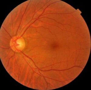
The Goldberg classification divides proliferative sickle cell retinopathy into five stages that follow its natural course.
It guides follow-up and treatment, since the stage predicts the risk of vitreous hemorrhage and detachment.
Knowing the stages helps clinicians to explain the disease to patients and to decide when to treat.
What the Goldberg Classification Describes
Goldberg described the progression in 1971 from peripheral arteriolar occlusion to retinal detachment.
The disease begins with sickled cells blocking peripheral vessels, and it proceeds through remodeling and new vessel growth.
Stages need not occur in every eye, and many eyes regress or stay stable (see sickle cell retinopathy).
The Five Stages of the Goldberg Classification
- Peripheral arteriolar occlusions mark stage I
- Peripheral arteriovenous anastomoses mark stage II
- Neovascular and fibrous proliferation, the sea fan, marks stage III
- Vitreous hemorrhage marks stage IV
- Tractional and combined retinal detachment mark stage V
Early stages are asymptomatic, and later stages cause floaters, flashes, and loss of vision.
Fundus Explorer Pro
Photograph the retinal findings described here with the phone already in your pocket — 22 D optics and built-in illumination in one handheld unit.
From Choroida — the team behind this siteSea Fan Neovascularization
Sea fans arise at the border between perfused and nonperfused retina.
They can autoinfarct and regress in many eyes, but they may also bleed or produce traction (see sea fan neovascular frond).
Genotype and the Goldberg Classification
Hemoglobin SC and sickle thalassemia carry a higher risk of proliferative disease than hemoglobin SS.
Downes and colleagues followed a cohort and reported the incidence and natural history of proliferative sickle retinopathy.
Spontaneous regression occurred in many eyes, so treatment decisions weigh risk and course.
Screening and the Goldberg Classification
Start dilated examinations in childhood, around age 10.
Repeat every one to two years, and more often with proliferative changes.
Wide-field imaging and OCT angiography help to find peripheral nonperfusion.
Treatment by Stage in the Goldberg Classification
- Observation and regular follow-up suit stages I and II
- Scatter laser or anti-VEGF may suit high-risk or bleeding eyes in stage III
- Observation for clearing, then laser or vitrectomy if blood persists, suits stage IV
- Vitrectomy with careful handling because of ischemia suits stage V
Surgery needs gentle technique and attention to anesthesia and oxygenation.
An Example Case
A 19-year-old man with hemoglobin SC disease has no symptoms and 20/20 vision.
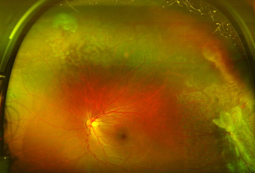
Wide-field imaging shows peripheral nonperfusion and a small sea fan in the superotemporal periphery, which is stage III.
He starts laser photocoagulation in the ischemic periphery, and the sea fan regresses over months.
At follow-up, there are no vitreous hemorrhages, and he keeps 20/20 vision.
Questions Patients Ask
Patients ask whether the disease will blind them, and most do not lose vision, though some are at risk without treatment.
They also ask whether the laser hurts, and anesthetic drops make treatment tolerable.
Because adherence matters, explain the need for yearly dilated exams.
Pitfalls to Avoid
- Starting screening too late in a child with sickle cell disease
- Treating every sea fan, although many regress by themselves
- Using high-pressure injection or gas bubbles without a plan for oxygenation
- Missing peripheral disease on a standard 30-degree photograph
Looking Ahead
OCT angiography and wide-field angiography reveal macular and peripheral ischemia earlier.
Also, gene therapy for sickle cell disease may change the course of retinopathy.
Therefore, ocular monitoring in treated patients remains important.
Practical Points
Dilate and examine the periphery with scleral depression or wide-field imaging.
Then record the stage using the Goldberg classification in each eye.
Also coordinate with hematology about hydration and transfusions before surgery.
Retinal Changes Beyond Neovascularization
Non-proliferative changes include salmon patch hemorrhages, iridescent spots, and black sunbursts.
Macular thinning may occur in the temporal macula, with loss of vision in some patients.
Because these signs mark ischemia, review them at each exam.
Anesthesia and Surgery Precautions
Avoid hypoxia, hypothermia, and acidosis during ocular surgery.
Use caution with epinephrine and with scleral buckling, which may reduce choroidal flow.
Then monitor for anterior segment ischemia.
Imaging for Staging
Wide-field fluorescein angiography shows peripheral nonperfusion, arteriovenous anastomoses, and neovascular fronds.
Also, OCT shows macular thinning in the temporal macula, which may reduce acuity.
Because staging depends on the periphery, use wide-field imaging whenever possible.
Counselling Patients and Families
Explain that most patients keep good vision with regular follow-up.
Then give a clear plan for exams and for urgent symptoms such as new floaters.
Because many patients are young, involve the family and the primary care team.
Summary
Stage each eye with the Goldberg classification, and follow the periphery.
Then treat high-risk sea fans, since many regress without treatment.
Because patients are young, plan lifelong follow-up.
Also coordinate with hematology at every step.


Document what you see
Two smartphone imaging tools built for everyday clinic use — one for the slit lamp, one for the fundus.
From Choroida — the team behind this siteReferences
- Goldberg MF. Classification and pathogenesis of proliferative sickle retinopathy. Am J Ophthalmol. 1971;71:649-665.
- Downes SM, Hambleton IR, Chuang EL, et al. Incidence and natural history of proliferative sickle cell retinopathy: observations from a cohort study. Ophthalmology. 2005;112:1869-1875.
- Salmon JF. Kanski’s Clinical Ophthalmology: A Systematic Approach. 9th ed. Elsevier; 2020.
- American Academy of Ophthalmology. Basic and Clinical Science Course, Section 12: Retina and Vitreous. AAO; current edition.

Writes and reviews the clinical articles on Educate. Every post is checked against peer-reviewed sources before it goes live.