Orbital emphysema is air trapped in the orbital soft tissues, usually after a fracture into a paranasal sinus.
Most cases look alarming, yet they resolve without intervention.
However, a few raise orbital pressure enough to threaten the optic nerve.
How Air Enters the Orbit
The thin medial wall and floor separate the orbit from the ethmoid and maxillary sinuses.
After a fracture, air moves into the orbit whenever sinus pressure rises.
In addition, a soft tissue flap may act as a one-way valve.
Nose Blowing and Orbital Emphysema
Patients often describe sudden lid swelling right after blowing the nose.
Because this history is typical, it should prompt a search for a wall fracture.
Swelling may appear days after injury, possibly because edema initially seals the defect.
All-fit Slit-Lamp Adapter
Record and share exactly what you see at the slit lamp. One adapter fits any slit lamp or surgical microscope — and any smartphone.
From Choroida — the team behind this siteOther Causes of Orbital Emphysema
Trauma accounts for most cases, although other routes exist.
For example, reports describe air entry after sinus surgery and dental procedures using compressed air.
Forceful nose blowing can occasionally cause it without any recognized injury.
What the Patient Reports
Lid swelling and fullness are the usual complaints.
Diplopia may follow, because air and edema restrict ocular movement.
Pain is often mild in uncomplicated cases.
Therefore, severe pain or falling vision should raise concern.
Crepitus in Orbital Emphysema
Gentle palpation of the lids may reveal a crackling sensation.
This sign reflects air within the subcutaneous tissue, and it strongly suggests a sinus communication.
However, purely deep orbital air may produce no crepitus.
Its absence therefore does not exclude the diagnosis.
Examining the Eye
First, record visual acuity, pupils, color vision, motility and intraocular pressure.
A relative afferent pupillary defect suggests optic nerve compromise and needs urgent attention.
Next, use the slit lamp to look for hyphema and signs of globe rupture.
Finally, inspect the disc and retinal circulation.
CT Findings in Orbital Emphysema
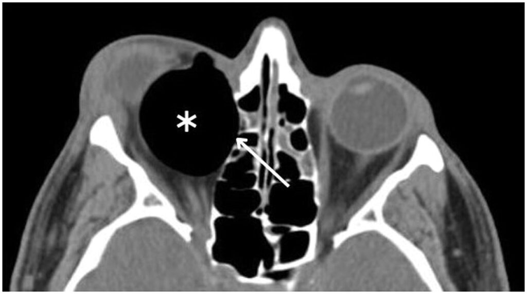
Noncontrast thin-section CT is the preferred study after orbital trauma.
Air appears as low-density pockets, often beside the fractured wall.
In addition, a tented posterior globe suggests dangerous tension.
Wood can mimic air, so review wide windows after penetrating injury.
Intraocular air points toward an open globe, while intracranial air suggests a skull base injury.
Differential Diagnosis
Retrobulbar hemorrhage also causes proptosis and raised pressure, although crepitus is not expected.
Preseptal hematoma causes marked lid swelling, yet it spares the deep orbit.
Orbital cellulitis evolves with fever and pain, while traumatic air appears abruptly (see orbital cellulitis).
Gas with fever and no trauma history suggests a gas-forming infection.
CT separates these conditions quickly.
Conservative Care for Orbital Emphysema
Uncomplicated air usually resorbs over days to a couple of weeks.
Management therefore centers on stopping further air entry.
Advise patients to avoid nose blowing and to sneeze with the mouth open.
Also discuss flying or diving with the treating team.
Antibiotics and Decongestants
Clinicians often prescribe oral antibiotics and nasal decongestants after a sinus-communicating fracture.
However, high-quality trials supporting either practice are limited.
Therefore, base the decision on sinus disease and local protocols.
Explain that these medications do not remove trapped air or relieve a compressive orbit.
Recognizing Compressive Orbital Emphysema
Vision-threatening compression is rare, but it can develop quickly.
Warning signs include increasing proptosis, a tense orbit, marked pressure elevation and a new afferent pupillary defect.
Vision may fluctuate as orbital pressure changes.
Retinal arterial pulsation or pallor also calls for immediate action.
Emergency Decompression
Treat suspected compression clinically, because waiting for imaging can cost vision.
Lateral canthotomy and inferior cantholysis reduce orbital pressure.
In addition, needle aspiration can release a discrete air pocket, although the evidence comes mainly from case reports.
Both procedures carry risks, so reassess vision and pressure afterward.
Fracture Repair After Orbital Emphysema
Air alone rarely decides whether a fracture needs surgery.
Instead, entrapment, diplopia, enophthalmos and defect size guide timing (see orbital blowout fracture).
Therefore, repeat the examination once air and edema settle.
Follow-Up and Counseling
Recheck vision, pupils, pressure and motility while swelling resolves.
Tell patients the crackling is usually temporary.
However, they should seek urgent care for reduced vision or increasing proptosis.
Pitfalls to Avoid
- Do not dismiss sudden swelling after nose blowing as an allergy
- Never rely on crepitus alone to judge orbital tension
- Do not delay decompression for CT when the optic nerve is failing
- Avoid assuming every low-density focus is air after penetrating trauma
- Never attribute gas to trauma when the patient appears septic


Document what you see
Two smartphone imaging tools built for everyday clinic use — one for the slit lamp, one for the fundus.
From Choroida — the team behind this siteReferences
- Zimmer-Galler IE, Bartley GB. Orbital emphysema: case reports and review of the literature. Mayo Clin Proc. 1994;69:115-121.
- Hunts JH, Patrinely JR, Holds JB, Anderson RL. Orbital emphysema: staging and acute management. Ophthalmology. 1994;101:960-966.
- Roelofs KA, Starks V, Yoon MK. Orbital emphysema: a case report and comprehensive review of the literature. Ophthalmic Plast Reconstr Surg. 2019;35:1-6.
- Carter KD, Nerad JA. Fluctuating visual loss secondary to orbital emphysema. Am J Ophthalmol. 1987;104:664-665.
- American Academy of Ophthalmology. Basic and Clinical Science Course, Section 7: Oculofacial Plastic and Orbital Surgery. San Francisco: AAO.

Writes and reviews the clinical articles on Educate. Every post is checked against peer-reviewed sources before it goes live.