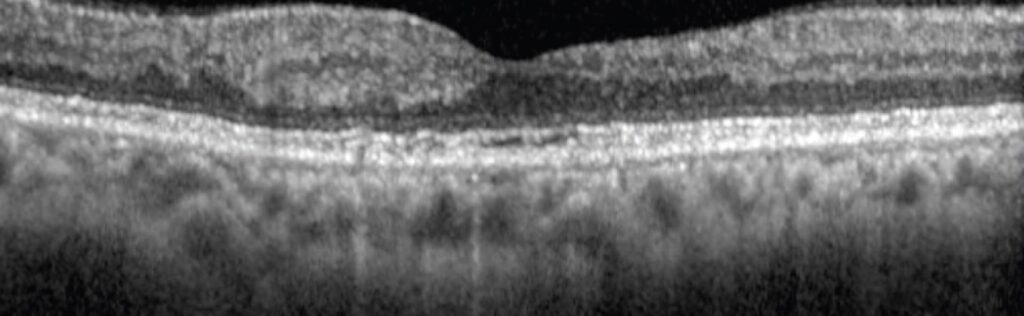
Disorganization of retinal inner layers, or DRIL, is an OCT sign in which the boundaries between inner retinal layers cannot be identified.
It predicts poor vision in eyes with diabetic macular edema and retinal vein occlusion, and it often persists after the edema resolves.
Recognizing DRIL helps clinicians to set realistic expectations before treatment.
What Disorganization of Retinal Inner Layers Means
DRIL describes loss of the normal boundaries between the ganglion cell layer, the inner plexiform layer, the inner nuclear layer, and the outer plexiform layer.
Sun and colleagues defined it as the inability to identify any of these boundaries over a horizontal length on a B-scan.
The change probably reflects damage to bipolar, amacrine, and horizontal cells that carry signals from photoreceptors to ganglion cells.
This loss disrupts the retinal pathway even when the photoreceptors survive.
How to Identify Disorganization of Retinal Inner Layers
Use a high-quality horizontal B-scan through the fovea.
Look for blurred or absent lines between the layers within the central 1 mm and beyond.
Measure the horizontal extent of the area, and record whether the disorganization is focal or wide.
Check several B-scans, since a single slice may miss a larger lesion (see ellipsoid zone disruption).
Fundus Explorer Pro
Photograph the retinal findings described here with the phone already in your pocket — 22 D optics and built-in illumination in one handheld unit.
From Choroida — the team behind this siteDisorganization of Retinal Inner Layers in Diabetic Macular Edema
Sun and colleagues found that DRIL predicted worse acuity at baseline and later visits.
A larger DRIL extent correlated with worse vision, and a change in DRIL over four months predicted a change in acuity.
Radwan and colleagues showed that DRIL after resolution of edema also related to final vision.
Das and colleagues linked inner retinal disorganization to outer retinal damage in a larger cohort (see diabetic macular edema).
Disorganization of Retinal Inner Layers in Other Diseases
- Branch retinal vein occlusion with macular edema
- Central retinal vein occlusion and macular ischemia
- Macular telangiectasia and other retinal vascular disease
- Retinal artery occlusion with inner retinal infarction
In vein occlusion, DRIL points to ischemic injury that may limit visual recovery (see branch retinal vein occlusion).
Using Disorganization of Retinal Inner Layers in Practice
Record the presence and extent of DRIL at baseline.
Counsel patients that vision may improve less than expected when DRIL is wide, even if the edema resolves.
Avoid over-treating eyes with stable DRIL and a dry macula, since further injections will not restore lost cells.
Use DRIL together with acuity, thickness, and ischemia on angiography.
Limits
Measurements vary between graders, and segmentation software cannot yet apply the definition reliably.
Poor image quality and macular fluid may hide boundaries.
Therefore, always review the B-scans manually.
An Example Case
A 60-year-old man has diabetic macular edema in his right eye, with acuity of 20/80 and a central thickness of 450 micrometers.
OCT shows a wide area of disorganized inner layers, with no clear boundaries over 1.5 mm.
After six injections, the edema resolves, but his acuity improves only to 20/60.
The persistent disorganization explains why vision lags behind the anatomic response.
Questions Patients Ask
Patients ask why vision stays blurred even though the swelling is gone, and the answer is that nerve cells in the retina may be damaged.
They also ask whether more injections will help, and extra injections do not repair lost cells.
Because the answer may disappoint, explain that early treatment protects vision and that low vision aids can help.
Pitfalls to Avoid
- Treating a dry macula with DRIL as a failure of therapy
- Mixing up DRIL with simple blurring from fluid
- Using one B-scan to judge a large area
- Forgetting to check for macular ischemia on angiography
Looking Ahead
Automated detection tools may measure DRIL extent and track change over time.
Also, researchers study whether early treatment lowers the risk of DRIL.
Therefore, DRIL may become a standard endpoint in trials.
Practical Points
Review the horizontal and vertical B-scans through the fovea.
Then record the DRIL extent in the chart, and compare with later scans.
Also discuss prognosis with the patient when DRIL covers the central region.
Tracking DRIL Over Time
Compare the horizontal extent at each visit.
Because DRIL may improve slightly after the edema resolves, repeat the scan at three and six months.
Then record the change in the chart.
Also share the findings with the patient, since they explain slow recovery.


Document what you see
Two smartphone imaging tools built for everyday clinic use — one for the slit lamp, one for the fundus.
From Choroida — the team behind this siteReferences
- Sun JK, Lin MM, Lammer J, et al. Disorganization of the retinal inner layers as a predictor of visual acuity in eyes with center-involved diabetic macular edema. JAMA Ophthalmol. 2014;132:1309-1316.
- Radwan SH, Soliman AZ, Tokarev J, et al. Association of disorganization of retinal inner layers with vision after resolution of center-involved diabetic macular edema. JAMA Ophthalmol. 2015;133:820-825.
- Das R, Spence G, Hogg RE, et al. Disorganization of inner retina and outer retinal morphology in diabetic macular edema. JAMA Ophthalmol. 2018;136:202-208.
- American Academy of Ophthalmology. Basic and Clinical Science Course, Section 12: Retina and Vitreous. AAO; current edition.

Writes and reviews the clinical articles on Educate. Every post is checked against peer-reviewed sources before it goes live.